 Open access
Abstract
The human body is colonized by a diverse community of microorganisms that are crucial for human health, while microbiota homeostasis is modulated by both environmental and host-derived cues. Specific microbial species form surface-attached microcolonies surrounded by their self-generated extracellular polymeric substances called biofilms. These complex assemblies may protect commensal bacteria, serving as a barrier against pathogenic microorganisms as well as hostile conditions, thus creating structuralized microbial communities for complex microbial interactions. However, biofilms formed by pathogenic bacteria will lead to invasion and perturbance of microbiota homeostasis, potentially leading to dysbiosis and diseases. In this review, we provide an in-depth analysis of the multiple-species biofilms on microbiota homeostasis within the human body. We describe the nature of biofilms in various anatomical regions, emphasizing their protective role against pathogenic invasions and their potential roles in causing disease under certain conditions. We also explore the microbial interactions between biofilms and microbial communities, emphasizing the significant potential of biofilms to alter the structure and composition of microbial assemblages. Finally, we suggest the directions for future biofilm research to enhance our understanding of the pathogenesis of diseases related to microbial dysbiosis and human health.
Microbial Communities

Biofilm Formation and Pathogenesis
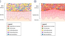
Microbial biofilms and the human skin microbiome
1 Introduction
The human body is composed not only of billions of human cells, but also in the unseen microscopic realm, it harbors a far greater number, ranging in the trillions, of microorganisms (Rackaityte et al. 2020). Various complex and diverse ecological microenvironments within the human body provide attachment points for these intricate microbial communities. These microbes assist in maintaining physiological homeostasis, preventing pathogenic invasions, and guiding human immune systems to uphold health and stability (Zhu et al. 2023). Increasing evidence showed that microbiota homeostasis is crucial for maintaining human health. The human microbiota is composed of a collection of dynamic microbial communities that inhabit various anatomical locations in the body. Accordingly, the coevolution of the microbiota within the host has resulted in these communities playing a profound role in promoting human health (Aggarwal et al. 2023; Hou et al. 2022). With the advent of metagenomics and next-generation sequencing technologies, new insights have been gained into the composition, structure, function, and dynamics of the microbiota at various body sites (Yuan et al. 2012; Brooks et al. 2015).
Microbes can typically exhibit two modes of growth: one involves free-floating, while the other entails attachment to solid surfaces through the formation of a biofilm, thereby establishing a spatial structure within the microbiota (Howlin et al. 2017; Liu et al. 2019). Biofilms comprise extracellular polymeric substances (EPS) produced by the microorganisms themselves, along with secreted extracellular polysaccharides, proteins, lipids, and nucleic acids (Zambrano et al. 2005). Biofilm formation is the outcome of physical and chemical interactions among microbial communities, serving as a conducive environment for microorganism survival and protection, as well as facilitating the aggregation and attachment of microorganisms (Jiang et al. 2018; Qu et al. 2020). Moreover, EPS provides a structurally-organized platform for microbes, facilitating the exchange of substances and the formation of symbiotic relationships among cells (Flemming et al. 2016). The formation of biofilms encompasses several stages: initial adhesion, microcolony formation, matrix production, and maturation, as well as maintenance and dispersion (Vlamakis et al. 2013; Sauer et al. 2004). In many parts of the human body, such as the oral cavity, intestines, and gastrointestinal tract, biofilm often form, composed of a diverse array of microorganisms (Hall-Stoodley et al. 2004), and these biofilms contain a variety of beneficial and harmful bacteria, intricately interacting within this complex microbial community (Shirtliff et al. 2002). Biofilms serve as reservoirs for microbes, releasing planktonic bacteria to microbial communities to maintain a stable microbial population. Current research on microbial community composition and species diversity often ignores the significant role of biofilms within these communities. More and more studies have revealed that biofilms are a crucial structural component in maintaining the stability of the human microbiota (Miller et al. 2021; Karygianni et al. 2020; Centurion et al. 2021), which provide a dynamic environment for microbial community assembly, where microbes can interact, communicate, and collaborate (Flemming et al. 2023; Nadell et al. 2016).
Microbiota homeostasis refers to a state where the composition and activity of a microbial community remain relatively stable under certain environmental conditions (Buchon et al. 2013). The interplay between biofilm and microbiota homeostasis is a complex and multifaceted relationship (Motta et al. 2021). The composition and behavior of microbiota can be influenced by the biofilm, while the characteristics of these communities can, in turn, affect the formation and stability of the biofilm (Gilbert et al. 2002; Wuertz et al. 2004). The biofilm is capable of maintaining the homeostasis of the microbiota on multiple levels, which is categorized into passive defense and active resistance. Several factors disrupt the homeostasis of the microbiota, such as the action of antibiotics and the invasion of bacteriophages. A study involving healthy human volunteers demonstrated that before, during, and following a five-day course of oral ciprofloxacin, there was a decrease in the diversity, richness, and evenness of fecal microbiota during the antibiotic treatment period (Dethlefsen 2008). Bacteriophages are considered to shape the taxonomic and functional composition as well as the stability of microbial communities. Research by Bao et al. (2018) indicates that treatments with lytic and temperate gut bacteriophages can regulate the composition and diversity of the gut microbiota. Lytic gut bacteriophages may promote a beneficial gut ecosystem, while temperate bacteriophages can foster conditions conducive to disease development (Bao et al. 2018). These disruptions can be overcome through the passive defense mechanisms of the biofilm. Furthermore, the biofilm enables the assembly of various components within the microbial community into higher-order structures. Within these structures, more complex interactions can occur, including quorum sensing, intra- and interspecies competition mediated by the type VI secretion system (T6SS), and horizontal gene transfer, all of which actively contribute to the maintenance of microbiota homeostasis.
Biofilms provide a certain level of passive defense capabilities for the microbiota. This protection encompasses not only tolerance to physical and chemical environmental factors but also extends to resistance against antibiotics and tolerance to interference by the human immune system (Davey et al. 2000). Microbial communities within biofilms typically exhibit relatively high antibiotic resistance. This is attributable to the biofilm's structure and extracellular matrix, which can limit the penetration of antibiotics, thereby reducing their bactericidal effectiveness (Costerton et al. 1999; Stacy et al. 2016). Additionally, certain microbes within biofilms may combat the effects of antibiotics by producing antibiotic-degrading enzymes or altering their metabolic pathways (Orazi 2019; Høiby et al. 2010). This antibiotic resistance often makes dominant microbes within biofilms more challenging to eradicate. Furthermore, microbial communities within biofilms can demonstrate tolerance to the human immune system (Yan et al. 2019; Rybtke et al. 2015). The structure and extracellular matrix of biofilms can mask microbial surface antigens, making it difficult for the host immune system to recognize and attack the microbes within the biofilm (Brady et al. 2007). Moreover, some microbes within biofilms may produce molecules that suppress immune responses, thereby reducing interference by the immune system (Roilides et al. 2015). This immune tolerance aids microbes in establishing persistent infections within the human body and maintaining the stability of the microbial community (Robinson et al. 2010). These protective mechanisms enable microbes within biofilms to survive and proliferate in complex and competitive environments, thereby maintaining the stability and functionality of the biofilm. By aiding dominant strains within the microbial community to consolidate their competitive advantage, these mechanisms influence the homeostasis of the microbial population. In turn, when external factors disrupt biofilms, the homeostasis of the microbial community can also change or become compromised. Such disturbances may originate from external sources, such as the use of antibiotics, changes in the nutritional environment, or interventions by the immune system (Logan et al. 2016). The overuse of antibiotics serves as a quintessential example (ChoWdhury et al. 2012). Antibiotics are typically designed to kill or inhibit the growth of bacteria, but they often indiscriminately affect both planktonic bacteria in the gut and microbes within biofilms (Ribeiro et al. 2020; Soares et al. 2012). This can lead to the disintegration of biofilms and the dissolution of microbial communities, disrupting the previously existing microbial homeostasis. When the homeostasis of the microbial community is altered or damaged, biofilms tend to release planktonic microbes, altering the balance within the organism. This disruption may result in the overgrowth of microbes or other microecological changes, potentially leading to infections, diseases, or immune responses.
Additionally, the unique environment of the biofilm enables the assembly of various components within the microbial community into higher-order structures, facilitating more complex interactions therein. The formation of biofilms starts with the adhesion of microbes, normally after receiving certain environmental cues. Once these microbes adhere to a solid surface and start aggregating into microcolonies, their growth and metabolic characteristics change. This aggregation fosters interactions among the microbes, including symbiosis and competition, thereby affecting the composition and functions of the microbial community (Hall-Stoodley et al. 2004; Flemming et al. 2019). Microbial biofilms typically exhibit complex structures, where porous architecture and negatively charged surfaces influence the spatial distribution of the microbes. For instance, biofilms display a characteristic vertical stratification, including the surface and base of the biofilm, as well as intermediate water channels. Near the surface, microbial density is higher due to it being the initial point of attachment. However, the bottom and water channels exhibit lower microbial density but facilitate the transport and distribution of substances (Rumbaugh et al. 2020; Flemming et al. 2010). The intricate architecture of biofilms leads to the formation of various complex microenvironments, which directly impact the interactions among the microbes (Arnaouteli et al. 2021). In some instances, the dense population within a biofilm intensifies competition for moisture and nutrients among the microbial community. In contrast, in other scenarios, it can strengthen mutually beneficial relationships aimed at enhancing surface colonization (Nadell et al. 2016; Steenackers et al. 2016). Moreover, within the microbial community of a biofilm, there exists a diversity of complex inter- and intra-species interactions, including quorum sensing and competition within the microbiota mediated by the T6SS. These complex interactions shape the microbial community and maintain microbial homeostasis.
Quorum sensing (QS) is a widely used cell-to-cell communication system by microbial communities, which plays a crucial role in the development and stabilization of microbial communities. QS is a process where microbes secrete and detect a variety of signaling molecules, such as autoinducing peptides and N-acyl homoserine lactones (AHLs). These molecules enable the microbes to sense and respond to cell density. Upon reaching a threshold concentration, these signaling molecules trigger alterations in gene expression, thereby synchronizing behaviors within the community (Hense et al. 2007). The dense microcolony connected with their self-generated channels within biofilms that provides an ideal environment for microorganisms to engage QS to coordinate social activities and influence the community's homeostasis (Mukherjee et al. 2019; Papenfort et al. 2016). For instance, QS facilitates swarming motility and coordinates microcolony formation at the early stage of biofilm formation, while during the maturation stage of biofilms, QS signaling is instrumental in regulating the biofilm's structure and stability (Whiteley et al. 2017). Furthermore, QS allows microorganisms to detect and respond to environmental changes, such as nutrient availability and host immune responses, thus maintaining stability in complex environments. Therefore, QS is also integral to the maintenance of microbial homeostasis.
Quorum sensing represents a social behavior among microbial populations, coordinated by the group, while the T6SS functions as a mechanism for mediating inter-bacterial killing. From a holistic perspective, the T6SS, prevalent in Gram-negative bacteria, aids these bacteria in establishing competitive advantages within the biofilm's microbiota and adapting to environmental stresses (Hood et al. 2010; Russell et al. 2014). Several types of protein secretion systems exist in Gram-negative bacteria, functioning to transport proteins to the extracellular environment and occasionally to adjacent prokaryotic or eukaryotic cells (Ho et al. 2014; Wang et al. 2021). The T6SS serves as a typical contact-dependent killing device. It is widely distributed among over 25% of Gram-negative bacteria. Morphologically and structurally resembling a contractile bacteriophage tail attached to the inner membrane of the cell, it constitutes a needle-shaped multiprotein machinery (Manera et al. 2022). Its structure comprises a membrane complex, a baseplate structure, and a sheath-tube complex. It is capable of delivering various effectors for interactions with bacteria within both biotic and abiotic environments (Bönemann et al. 2010; Yu et al. 2021). Recently, the involvement of the T6SS has been demonstrated in various interactions within the microbiota, ranging from bactericidal activities to intra- and interspecies competition within mixed-culture biofilms (Schwarz et al. 2010; Ren et al. 2023). Within the biofilm, a microbiota-dense spatial environment, interactions mediated by the T6SS are more frequent and efficient. Through intense competition for colonization, they alter the structure and composition of the microbiota, thereby influencing its homeostasis.
Overall, biofilms play a dual-edged role in microbial community stability, enhancing the prospering of beneficial constituents while exacerbating the deterioration of detrimental ones. On the one hand, in terms of sustaining microbiota homeostasis, biofilms contribute to reinforcing the dominant position of resident microbes and blocking the invasion of external microbial species (Lepargneur and Rousseau 2002). On the other hand, certain bacterial pathogens (such as Pseudomonas aeruginosa), can invade the microbiota by forming strong biofilms and potentially causing primary or secondary infections, particularly when host immunity is compromised (Schulze et al. 2021; Lebeaux et al. 2013). In certain infection scenarios such as cystic fibrosis, microbiota homeostasis is completely disrupted by biofilms dominated by pathogenic microbes. These pathogen-dominated biofilms are stubborn, difficult to eradicate, and pose a serious threat to human health. Therefore, biofilms play a crucial role in the maintenance of microbial homeostasis.
In this paper, we present a comprehensive review based on recent reports, focusing on the relationship between human biological membranes and the homeostasis of microbiota across four distinct anatomical regions: the gastrointestinal biofilm, oral and dental biofilm, respiratory tract biofilm, and reproductive tract biofilm. The review analytically emphasizes various aspects depending on the microbiome stability and disease occurrence in different anatomical locations, as well as the varying focal points of current research interests.
2 The impact of gut biofilms on microbial community homeostasis
The intestine is one of the most crucial "organs" in the human body, characterized by a large and diverse gut microbiota (Sommer et al. 2013). This microbiota can interact with the host through the intestinal mucosa, playing significant roles in digestion, immunity, and nutrition (Tremaroli et al. 2012). Gut microbes inhabit various parts of the human intestine, forming a dynamically changing community (Yang 2020). The physiological composition of the gut microbiota also exhibits temporal and spatial specificity (Mallott et al. 2021; Lotstedt 2023). With the application of molecular biology techniques in the study of gut microbiota, it has been confirmed that these microbes not only degrade indigestible nutrients in food and provide various nutrients to the human body but also promote the differentiation and maturation of intestinal epithelial cells, activate the intestinal immune system, resist the invasion of foreign microbes, maintain the function of the intestinal barrier, and regulate the storage and metabolism of human energy (Ratiner 2023; Donald et al. 2023; Mazmanian et al. 2005). The relationship between gut microbiota and human diseases is currently receiving considerable attention. For simplicity and convenience, most studies have focused on the fecal microbiome, neglecting the spatial organization of intestinal communities, such as those associated with the intestinal lumen, food particles, or mucosa. Consequently, intestinal biofilms have received limited attention and are primarily associated with disease contexts (de Vos and de Vos 2015). However, biofilms or biofilm-like structures in the human gut may significantly impact the functionality of the gut microbiome and its interactions with the host. How these processes are regulated in gut symbionts is largely unknown.
The physiological diversity of the intestinal microbiota contributes to the maintenance of stability within the entire microbial community. The intestine is the most densely populated and diverse organ for microbial colonization, and the gut microbiome is highly complex, containing over 10 million genes (de Vos and de Vos 2015; Lahti et al. 2014; Li et al. 2014). The predominant bacterial phyla in the gut are Firmicutes and Bacteroidetes, accounting for 90% of the gut microbiota. Other phyla present in the intestinal environment include Actinobacteria, Proteobacteria, Clostridia, and Verrucomicrobia (Arumugam et al. 2011). Notably, under Firmicutes, there are 200 different genera, including Bacillus, Lactobacillus, Enterococcus, Clostridium, and Ruminococcus. While lactic acid bacteria are beneficial for health, some genera of Firmicutes, such as Staphylococcus aureus and Clostridium perfringens, are harmful when they overgrow (Arumugam et al. 2011). Dominant genera within the Bacteroidetes phylum are Bacteroides and Prevotella. Less abundant, Actinobacteria are primarily represented by Bifidobacterium, known for its positive impact on health. Under Proteobacteria, some well-known pathogens include Escherichia coli, Helicobacter pylori, Shigella, Salmonella, and Campylobacter (Aggarwal et al. 2023). Although the composition of the gut microbiota varies during different stages of an individual's life cycle, overall, the composition of the gut microbiota is in a state of dynamic equilibrium.
Intestinal multi-microbial biofilm and host mucous layer Biofilm maintains microflora homeostasis by resisting the invasion of pathogenic microorganisms. Hosts employ multiple mechanisms to maintain intestinal homeostasis and prevent aberrant immune responses against the microbiota. One such protective mechanism involves the mucus layer covering the surface of the colonic epithelium, serving as a barrier to separate epithelial cells from intestinal microbes and prevent the penetration of microbes into the epithelial cells (Tropini et al. 2017). This mucus layer prevents pathogens from reaching and residing on the intestinal epithelial surface, thereby forming an essential component of host immunity, and the origin of interactions between this region and bacteria is ancient and evolutionarily conserved (Johansson et al. 2014; Donaldson et al. 2016). This layer is constantly renewed and acts as a trap for both commensal residents and pathogens, preventing their penetration (Johansson et al. 2011). Several indications suggest that gut microbes reside in the colonic mucus in the form of a multi-microbial biofilm, an aggregation of microbes enclosed in an extracellular matrix (Sonnenburg et al. 2004). Bacteria within the biofilm cooperate or compete with each other, forming a complex community that plays a key role in maintaining the stability of the microbiota (Tytgat et al. 2019). In summary, a healthy intestinal biofilm is beneficial to the host as it participates in the exchange of nutrients on the epithelial surface, increases colonization resistance, and protects the host from invasion by intestinal pathogens (Sonnenburg et al. 2004; Hooper et al. 2001). Disruption of a healthy mucosal biofilm leads to the proximity or direct contact of pathogenic invasive biofilms with colonic epithelial cells, thereby causing inflammation (Dejea et al. 2018).
In a healthy physiological state, it is generally observed that the intestinal microbial biofilm and the host mucosal layer biofilm provide a physical barrier for the microorganisms. This barrier offers protection against antibiotics, digestive enzymes, and the host's immune system attacks. This phenomenon is of significant importance for maintaining the homeostasis of the host's intestinal environment (Rinninella 2019) (Fig. 1B). One of the most important is the microbiome's resistance to colonization by invasive pathogens, with invasive species' bacteria suppressed from colonizing the gut because they cannot replace native species that are already established (Donaldson et al. 2016). The ability of bacteria to colonize the gut is determined by their ability to utilize specific limiting nutrients. In intestinal microbiota species that have reached a stable state, the biome has difficulty in vacating available nutrient niches, which is a natural rejection of external invading microorganisms. However, when the healthy intestinal biofilm is exposed to antibiotic overuse and physical damage, the original homeostasis is readily disrupted, leading to a weakened defense mechanism. Consequently, the intestinal epithelial cells become exposed to the intestinal contents, increasing their susceptibility to invasion by pathogenic microorganisms (Fig. 1A). In a model of intestinal niche competition, the two commensals, Escherichia coli HS and Nissle 1917, successfully inhibited the colonization of Escherichia coli EDL933 (Ibekwe 1917). This phenomenon, known as the "barrier effect", is predicted by Freter's nutrient-niche hypothesis and is referred to as colonization resistance (CR) (Hentges 1984; Stecher et al. 2011; Jiang et al. 2021; He et al. 2014). The repulsion of foreign invading microorganisms is often mediated by material factors. For instance, the engineered Escherichia coli Nissle 1917 in the gut achieves resistance to the invasion of Salmonella Typhimurium through ecological sequestration of iron. After a single dose, the levels of Salmonella Typhimurium during infection are reduced by more than 2 log units through the restriction of iron availability. Utilizing Escherichia coli Nissle 1917 before Salmonella Typhimurium infection can decrease the colonization rate by 445 times (Deriu et al. 2013). Another quintessential example is that of probiotics preventing the invasion and colonization of external pathogens by forming biofilms. Hooper et al. reported that the presence of probiotic biofilms in a healthy gut can prolong the residence time of bacteria, facilitating the exchange of nutrients between the host and the microbial community (Hooper et al. 2001). Research by Halfvarson et al. found that probiotics can achieve permanent colonization in the host mucosa through biofilm formation, thereby preventing the colonization of pathogens (Halfvarson 2017). Another intriguing example is the discovery of a specific C-type lectin, named CTL33, in the gut of the invertebrate black tiger shrimp. CTL33 facilitates the establishment of the gut microbiota within the intestinal tract by mediating biofilm formation, thereby maintaining the homeostasis of the gut microbial community (Harrell et al. 2020).
The intestinal microbial biofilm facilitates communication among microorganisms, where interactions mediated by chemical signaling contribute to the regulation of the behavior and composition of the microbial community. One of the well-studied cell-to-cell communication mechanisms is quorum sensing. Chemical signaling through quorum sensing is involved in regulating the bacterial group behaviors, often required for host colonization (Oliveira et al. 2023). Recent studies have indicated that a significant proportion of gut-associated bacteria, capable of encoding the AI-2 synthase (LuxS) or producing AI-2, have been identified in the Firmicutes and Proteobacteria phyla, as well as in some Bacteroidetes and Actinobacteria (Antunes et al. 2005; Lukáš et al. 2008). AI-2 can shape the composition of the murine gut microbiome by influencing the most abundant phyla in the intestine—Bacteroidetes and Firmicutes (Lukáš et al. 2008). In addition, AI-2 produced by Clostridium difficile is involved in regulating species interactions, specifically by affecting the multispecies biofilms formed with fragile Bacteroidetes. These bacteria respond to AI-2 produced by C. difficile, which induces metabolic reactions that subsequently inhibit the growth, biofilm formation, and virulence of C. difficile (Slater et al. 2019). Furthermore, the detection of AHL molecules in the murine gut suggests their production by members of the gut microbiota, although the specific members remain unidentified. Pathogens like small intestinal bacterial overgrowth produce AHL in the gut, activating the AHL receptor SdiA in Salmonella Typhimurium (Swearingen et al. 2013). Probiotics such as Bacillus subtilis and Lactobacillus plantarum can inhibit the peptide-based quorum sensing system of Staphylococcus aureus, promoting its decolonization (Dyszel et al. 2010).
Apart from quorum sensing, the T6SS serves as crucial antibacterial weaponry in regulating interactions among mammalian gut bacteria. The mammalian gut harbors the densest microbial communities and the most extensively formed natural biofilm structures. Within this densely populated spatial environment where space and nutrients are limited, microbial competition for resources intensifies. In such a dense environment, the T6SS, functioning as a contact-dependent killing apparatus, operates more frequently (Wexler et al. 2016; Russell et al. 2014). Wexler et al., through their investigation of the gut commensal bacterium Bacteroides fragilis, established that there are over 10 (Zambrano et al. 2005)T6SS firing events per gram of colonic contents per minute. Moreover, these microorganisms require their T6SS to continuously function within the gut (Wexler et al. 2016). Translation: Additionally, among the analyzed 205 human Bacteroidetesgenomes, 130 T6SS loci were identified, indicating that approximately one-fourth of the human gut microbiota encodes at least one T6SS (Coyne et al. 2016). The T6SS is not only crucial in shaping microbial community composition but may also be a major determinant of host gut microbiota composition. Human gut commensal B. fragilis effectively kills T6SS-dependent human gut commensal Bacteroidetes in vitro through a T6S-dependent mechanism (Russell et al. 2014). Verster et al. (2017) employed metagenomic analysis to delineate the distribution and function of the three sets of T6SS (GA1-3) within the Bacteroides genus in the human gut. They confirmed that B. fragilis GA3 mediates robust inter-strain competition, which correlates with an increase in the abundance of Bacteroides species. This suggests that the system confers a competitive advantage within Bacteroides-rich ecosystems (Coyne et al. 2016). Furthermore, T6SS plays a significant role in controlling interactions between the microbiota and invasive pathogens, thereby profoundly influencing host health. Enterotoxigenic B. fragilis (ETBF), which produces the B. fragilis toxin (BFT), can cause acute diarrhea and is associated with active inflammatory bowel disease (IBD), advanced colorectal cancer, and sepsis. Conversely, non-enterotoxigenic B. fragilis (NTBF) strains are commensal and can protect the host from inflammatory diseases (Wu et al. 2009; Choi et al. 2016; Mazmanian et al. 2008). Hecht et al. (2016) demonstrated that the commensal bacterium NTBF utilizes T6SS to resist colonization by the ETBF pathogen, thereby limiting toxin exposure. T6SS also serves as a critical weapon for many pathogenic microorganisms to invade, colonize, and disrupt microbiota homeostasis. Vibrio cholerae poses an increasingly serious threat to global health, and its T6SS is functional, mediating antagonistic interactions between bacteria during infection (Fu et al. 2013; Brooks et al. 2013).
The discussion above pertains to healthy physiological states. However, in the extreme scenario of severe infection, it is noteworthy that when the homeostasis of the microbiome is disrupted, certain invading pathogenic microorganisms often can reshape the composition and function of the microbiota through their robust biofilm-forming capabilities, leading to numerous intractable clinical issues. These include latent bacterial infections, unculturable microbes, persistent cells, and multi-drug tolerance in biofilms (Lewis and Lewis 2007) (Fig. 1C). Typical invading pathogens with strong biofilm-forming abilities include Helicobacter pylori, Salmonella spp., and enteropathogenic Escherichia coli. The persistence of Helicobacter pylori in human infections and its resistance to commonly used antimicrobial treatments are attributed not only to genetic variations but also to its ability to form biofilms, a strategy to overcome environmental stress and protect itself (Cammarota et al. 2012). Coticchia et al. used endoscopically guided biopsies and scanning electron microscopy to quantify the density of Helicobacter pylori biofilms on human gastric mucosa, observing significant alterations in the intestinal biofilm composition relative to a healthy physiological state (Coticchia et al. 2006). Fusco et al. reported that in conditions of microbial dysbiosis, the probiotic state of intestinal microbial biofilms changes, leading to a loss of protective capabilities, the occurrence of opportunistic infections, and the frequent emergence of multi-drug resistant biofilms of Pseudomonas aeruginosa and Staphylococcus aureus (Fusco et al. 2021). Sherlock et al. reported enhanced biofilm-forming abilities in intestinal invasion by certain enteropathogenic Escherichia coli strains, mediated by the potent bacterial adhesin AIDA, which aggregates bacteria and colonizes biofilms, disrupting healthy microbial homeostasis (Sherlock et al. 2004). Furthermore, Harrell reported that chronic infections caused by biofilms of Salmonella Typhi and Salmonella Typhimurium typically have high levels of bacterial replication and associated pathogen burden. Biofilms alter the growth physiology of bacteria, increasing their tolerance to antibiotics and long-term exposure, and developing tolerance against other host responses such as the complement system, antimicrobial peptides, antibodies, and the phagocytic activities of neutrophils and macrophages, ultimately benefiting the pathogen. Chronic inflammation caused by biofilms of Salmonella Typhi and Salmonella Typhimuriumis also associated with several malignancies related to chronic infections (Wessman et al. 2015), including gastric adenocarcinoma and lymphoma caused by Helicobacter pylori infection, or gallbladder cancer caused by Salmonella Typhi infection (Harrell et al. 2020). In summary, aberrant biofilm characteristics are associated with gastrointestinal diseases (Motta et al. 2021). Furthermore, characterizing the changes in biofilms and conducting causative studies are essential to elucidate their role in pathophysiology and to provide options for therapeutic intervention.
3 Oral biofilm and microbiome homeostasis: a balance susceptible to lifestyle disruption
The human oral microbiome is highly diverse and comprises a complex ecosystem, including bacteria, microeukaryotes, archaea, and viruses (Belibasakis and Belibasakis 2018). These communities display intricate and highly structured biogeography, shaping metabolic exchanges on a local scale and arising from various microenvironments present in the oral cavity (Baker 2023). The oral microbiome colonizes five major physically distinct niches: saliva, the tongue, oral mucosa, mineralized tooth surfaces, and periodontal tissues, each harboring distinct microbial assemblages (Caruso et al. 2020). The accessibility of the oral microbiome enables direct capture of biofilm formation and community assembly processes at sites of interest, thereby offering a powerful model system for exploring and understanding complex microbiomes (Jiao et al. 2019). Biofilms formed on the surface of teeth or dental materials, known as oral biofilms, are often considered virulence factors in many oral infectious diseases, including caries, periodontitis, and endodontic infections. Complex dynamic interactions between microbes, the host, and the diet lead to microbial colonization and the formation of oral biofilms (Marsh et al. 2017).The composition of the microbiome is influenced by the oral environment, with changes in local environmental conditions, especially dietary habits, affecting microbial interactions within these oral communities and determining, to some extent, whether the relationship between the oral microbiome and the host is symbiotic or potentially destructive (dysbiosis), thereby increasing the risk of diseases such as caries and periodontal disease (Roberts et al. 2015; Marsh 2003).The homeostasis of the oral microbiome is also associated with systemic diseases such as type 2 diabetes, cardiovascular diseases, cancer, preterm birth, and Alzheimer’s disease, but the molecular mechanisms supporting any direct role of the oral microbiome in systemic diseases remain to be elucidated (Eckburg et al. 2005; Oliveira et al. 2020).
The oral microbiome biofilm and the stability of the oral microbial community are both influenced by lifestyle, particularly dietary habits. The stability of the oral microbiome refers to a state of relative balance within this complex ecosystem, where microbes are neither causing disease nor completely eradicated by the immune system, representing a dynamic equilibrium that changes with the host's age. Neonates are born with nearly sterile oral cavities, but they are quickly colonized by environmental microbes, resulting in a relatively simple oral microbiome at this stage. Research by Tuominen et al. found that the neonatal oral microbiome is not significantly modulated by the birth canal or maternal oral microbiome but is notably associated with the microbiota in the placenta, suggesting a prenatal origin for the neonatal oral microbiome. (Tuominen et al. 2019) During this process, different bacterial species interact physically and metabolically, forming the initial biofilm community structure. Subsequently, the eruption and exfoliation of teeth provide new, non-shedding surfaces for colonization by commensals and opportunistic pathogens, leading to changes in the oral microbiome biofilm and microbial community. Additionally, the oral microbiome biofilm is influenced not only by age, diet, oral hygiene, and immune status but also by certain medications, such as antibiotics and drugs that reduce saliva secretion (Bowen et al. 2018). The key role of diet in microbial colonization has been well demonstrated in patients or experimental animals. Dental caries is a multi-microbial disease, with different species associated with various stages of the disease, including non-mutans Streptococci, Actinomyces, Bifidobacterium, Lactobacillus, Propionibacterium, Veillonella, and Treponema (Gross et al. 2010). When the host is excessively exposed to dietary sugars, the structure and composition of the biofilm formed on teeth undergo significant changes, with the residing microbial community becoming highly adapted to metabolizing carbohydrates and producing acids that cause caries. Oral pathogens, particularly Streptococcus mutans, can work synergistically with other acid-producing bacteria to induce ecological changes in the biofilm, disrupting the original microbial community homeostasis and forming a biofilm community, structure, and metabolism conducive to caries development (Bowen et al. 2018; Takahashi et al. 2011). However, during this process, some microbial interactions are beneficial, disrupting cariogenic pathogens. When the host's diet is not rich in fermentable carbohydrates, especially sucrose, commensals have a significant advantage over cariogenic pathogens (Bowen et al. 2018). Many commensals associated with oral health can adhere more strongly to saliva-coated tooth surfaces, grow better than S. mutans and many other acidogenic bacteria, and have multiple mechanisms to disrupt their establishment and growth. The dynamic balance between commensals and opportunistic pathogens may be disrupted by frequent sugar intake and poor oral care, promoting the development of deleterious biofilms near tooth surfaces (Dental 2017). The formation of caries can affect individuals of any age and involves interactions between teeth, microbial biofilms on tooth surfaces, and dietary sugars (Adler et al. 2013). With the continuous increase in dietary sugar, the incidence of caries is also rising (Van Dyke and Van Dyke 2017). Furthermore, poor oral care habits often disrupt the healthy homeostasis of the oral microbiome, inducing periodontitis. Periodontal disease (PD) is a complex infectious disease that starts with the disruption of microbial community homeostasis. This disease induces a host inflammatory response, leading to damage to the soft tissues and connective tissue tooth-supporting structures. In advanced cases, it also leads to tooth loss (Van Dyke and Van Dyke 2017; Greenwood et al. 2020). The etiology of PD is related to microbial community dysbiosis caused by poor oral care habits and dietary practices, with microbes, genetic susceptibility, and lifestyle determining the disease's development and severity (Fig. 2) (Heidari and Heidari 2014; Heidari et al. 2019).
Analysis of dental biofilms (plaque) in their natural state in clinical samples has revealed that the structure and composition of dental plaque are much more complex than previously thought (Simon-Soro et al. 2022). It contains clusters of spatial heterogeneity within the dense ecological space of the biofilm, where interactions within the oral microbiome and between the oral microbiome and the host are intricate. These complex interactions play a crucial role in maintaining microbial community homeostasis, including physical interactions and those mediated by chemical signaling molecules. Porphyromonas gingivalis has been observed to engage in direct physical interactions with many different members of the oral community, including early and late colonizers (Diaz et al. 2020). Studies by Ng et al. have shown that co-aggregation of P. gingivalis with Streptococcus gordonii is mediated by the fimbrial protein FimA, which specifically binds to glyceraldehyde-3-phosphate dehydrogenase (GAPDH) located on the surface of S. gordonii (Ng et al. 2016). Research by Zhu et al. indicates that the gingipains RgpA, RgpB, and Kgp of P. gingivalis play a significant role in vitro in the synergistic biofilm formation with Treponema denticola (Zhu et al. 2013). These direct contact interactions enhance the competitive advantage of specific colonizing species within the oral microbiome, affecting microbial community homeostasis by altering biofilm structure. Furthermore, chemical signaling-mediated interactions within the biofilm are more complex, with a network of synergistic and antagonistic interactions in the oral biofilm leading to new properties of persistence, stability, and spatial structure. These characteristics mediate the ecological shift from health to oral disease (Diaz et al. 2020). Wang et al. have demonstrated that the production of the quorum sensing signal molecule AI-2 in mixed biofilms of Streptococcus mutans and S. gordonii affects the structure, volume, and species composition of the symbiotic biofilm between these closely related oral bacteria, promoting co-aggregation of mixed species (Wang et al. 2017). From this, it can be inferred that in such multi-microbial biofilms, the QS signal molecule AI-2 consolidates the ecological position of the pathogen S. mutans, accelerating the dysbiosis of the oral microbiome. Understanding the fundamental characteristics of the interaction networks within oral microbial biofilms not only aids in the prevention of oral diseases but also contributes to the development of effective strategies to manipulate oral communities.
4 Biofilms present significant challenges in the treatment of respiratory infections
In a healthy state, the microbial communities in the oral cavity and respiratory tract exhibit a high degree of shared composition and similarity, including genera such as Corynebacterium spp., Staphylococcus spp., Moraxella, Haemophilus, and Streptococcus, among others (Ramakrishnan et al. 2016; Dickson et al. 2015). However, research on respiratory tract biofilms has largely focused on persistent and intractable chronic infections initiated by biofilms. Therefore, this section will primarily discuss the impact of biofilms on the stability of microbial communities in respiratory tract infections.
In chronic infections, notorious pathogens such as P. aeruginosa, Acinetobacter baumannii, Staphylococcus aureus, and the fungus Candida albicans often achieve effective invasion through their robust virulence and rapidly colonize by forming strong biofilms (Dickson et al. 2015; Guo et al. 2022; Wang et al. 2022; Ost et al. 2021; Chua et al. 2016). They dominate ecological niches through intra- and inter-species competition, ultimately reshaping the composition and function of the microbial community. A case in point is the biofilm infection of P. aeruginosa in the respiratory tract. P. aeruginosa, a common pathogen in nosocomial infections, often causes infections in immunocompromised individuals, particularly in Intensive Care Units (ICU) and respiratory patients, and frequently exhibits multidrug resistance (Qin et al. 2022). Its formidable biofilm-forming capability poses significant challenges in clinical treatment, making it one of the multidrug-resistant ESKAPE pathogens, which include Enterococcus, S. aureus, K. pneumoniae, A. baumannii, P. aeruginosa, and E. coli (Daikos et al. 2021). P. aeruginosais prevalent in respiratory infections, accounting for 16%–40% of ventilator-associated pneumonia (VAP) cases and can cause chronic or recurrent infections in CF or bronchiectasis patients (Sadikot et al. 2005; Finnan et al. 2004; Babich et al. 2020). The invasion and colonization process of P. aeruginosa, along with the development of its biofilm, changes the microbial community, regulated by the bacterial intracellular signaling molecule c-di-GMP, coordinating the transition from motility to a sessile lifestyle (Sauer et al. 2022; Jenal et al. 2017; Chua et al. 2014). The correlation between high intracellular c-di-GMP concentrations and biofilm formation or low c-di-GMP levels and motility has been confirmed in several bacteria, including P. aeruginosa and E. coli (Basu Roy et al. 2014). Initially, the invasion process is associated with low levels of intracellular c-di-GMP, corresponding to enhanced virulence and invasiveness of P. aeruginosa, allowing it to breach the barriers of the host and other competitors (Chua et al. 2014). This process represents a challenge to the homeostasis of the microbial community in a healthy physiological state. As native microbes are overthrown and the host's immune defenses fail, planktonic cells gradually adhere to tissue surfaces, divide, and produce extracellular polymer (EPS). In successful infection scenarios, biofilms develop into three-dimensional, surface-attached aggregates (Sauer et al. 2022). This marks the complete disruption of the normal microbial community homeostasis, with drastic changes in microbial composition and structure, and the establishment of a "new order" within the microbial community. P. aeruginosais, a typical example of biofilm impact on microbial communities, as its biofilms often develop into more complex mushroom-like, columnar structures, with bacterial cells growing into more complex multicellular mature forms and showing differentiation (Wood et al. 2000; Petrova et al. 2009). The "new steady-state order" includes intra- and inter-species competition driven by cell crowding, chemical gradients, and nutrient competition, as well as group communication mediated by bacterial quorum sensing systems, cooperative resistance to antibiotics, and frequent horizontal gene transfer within the biofilm (Costerton et al. 1999; Nadell et al. 2016; Papenfort et al. 2016; Stewart et al. 2008; Madsen et al. 2012). At this point, the microbial community composition under the domination of pathogenic biofilms becomes more uniform, converging towards a new "steady state," characterized by microbial community polarization, often accompanied by host immune stress, inflammatory storms, and the emergence of secondary infections.
The aforementioned scenario presents an idealized simple model. However, in reality, respiratory infections are often not caused by a single pathogen. Instead, they frequently involve co-infections and secondary infections involving various pathogens such as viruses with bacteria, bacteria with bacteria, viruses with fungi, and bacteria with fungi. According to a report from the UK, 60% of patients with CF were found to harbor Aspergillus species, with co-infections of P. aeruginosa and Aspergillusin CF patients, about half of whom were children (Hughes et al. 2022). In ICU, mixed microbial biofilms are commonly observed, such as clinically common biofilms comprising P. aeruginosa and Staphylococcus aureus (Wang et al. 2022; Jabra-Rizk et al. 2006). Extended mechanical ventilation in ICU, which is a significant risk factor for nosocomial infections, exacerbates this issue (Cimolai and Cimolai 2021). A study from Russia in March 2020 on COVID-19 found that 433 cases (35.96%) of COVID-19 patients tested positive for various bacterial pathogens, with infections from S. pneumoniae, S. aureus, and Haemophilus influenzae constituting the majority of secondary pneumonia cases, making secondary bacterial pneumonia a particularly dangerous complication (Sharov and Sharov 2020). In disease settings, multiple microbial infections often result in worse outcomes than single-species infections, and the impact of biofilms from multiple microbial infections on microbial community homeostasis is more complex, with numerous studies describing cross-kingdom interactions that alter biofilm structure (Weinstein et al. 1983; Pammi et al. 2014). Notably, the presence of P. aeruginosa and Burkholderia cepacia complex, pathogens commonly isolated in adult CF patients, significantly affects the composition and presence of other species (Zemanick et al. 2017; Stressmann 2012). The presence of either of these organisms is associated with a lower likelihood of future infections with methicillin-sensitive S. aureus or S. maltophilia (Granchelli 2018). These pathogens are thought to directly cause a decrease in microbial diversity, as they can compete with other microbes found in the lungs of CF patients, limiting the acquisition of other pathogens and controlling pre-established microbial communities (Welp et al. 2020).
In respiratory infection biofilms, complex interactions exist among various pathogenic microorganisms, including direct physical contacts and interactions mediated by chemical signaling molecules. These intricate interaction networks impact the stability of microbial communities. As a key component of biofilms, changes in the EPS matrix structure are considered highly associated with alterations in biofilm structure. A clinical model of dual biofilm comprising P. aeruginosa and Acinetobacter baumannii revealed that the extracellular polysaccharide Psl and eDNA of PAO1 play a crucial role in spatial interactions, with the presence of A. baumannii R2 protecting the antibiotic-sensitive P. aeruginosa PAO1 in the dual-species biofilm (Wang et al. 2022; Pammi et al. 2014). A notable cross-kingdom interaction is the collaborative biofilm formation of Staphylococcus aureus with the fungus Candida albicans, particularly where S. aureus can adhere to the fungal hyphae of C. albicans. This mixed-species biofilm can protect S. aureus from the effects of miconazole and high doses of vancomycin, exhibiting dependence on eDNA and β-1,3-glucan (Harriott et al. 2009; Kean et al. 2017). Chemical signaling-mediated interactions among various pathogenic microorganisms within the biofilm are even more complex. A quintessential example is the QS signaling system among microbial communities. The small molecule HQNO secreted by P. aeruginosa has been shown to have a profound impact on the physiology of S. aureus in vitro, protecting S. aureus biofilms from the effects of cell wall-targeting antibiotics, including the frontline drug vancomycin and various β-lactam antibiotics (Orazi 2017). Conversely, the quorum-sensing signal farnesol from C. albicans can disrupt the integrity of the S. aureus cell membrane and alter the drug sensitivity of S. aureus (Jabra-Rizk et al. 2006; Inoue et al. 2016). Horizontal gene transfer (HGT) associated with biofilm formation and development is highly related to the adaptation and evolution of bacteria (Madsen et al. 2012). HGT intertwines microbial communities within biofilm communities from the genetic to the community level through complex interactions (Burmølle et al. 2006). HGT in bacteria can occur through three different mechanisms: direct cell-cell contact (conjugation), phage-mediated DNA transfer (transduction), and uptake of naked DNA by competent cells (transformation) (Madsen et al. 2012). A typical example is the transfer of biofilm-associated plasmids among bacteria through conjugation. Many of the aforementioned examples of biofilm-enabling plasmids originate from E. coli, but biofilm priming encoded on plasmids has been identified in both Gram-negative and Gram-positive bacteria such as the Pseudomonas putida TOL plasmid, Lactococcus lactis pAMb1, Azospirillum brasilense plasmids and the Enterococcus faecialis pBEE99 (D'Alvise et al. 2010; Luo et al. 2005; Petrova et al. 2010). These plasmids can promote biofilm formation in microbial communities, with knockout studies of the conjugative plasmid pBEE99 in Enterococcus faecalis showing a 70% reduction in biofilm formation in the parent strain (Coburn et al. 2010).
Dysbiosis of the microbial community caused by exogenous pathogenic biofilm infections often leads to numerous intractable respiratory infections. CF, a hereditary disease, is caused by mutations in the cystic fibrosis transmembrane conductance regulator (CFTR), an ion channel primarily responsible for regulating chloride and bicarbonate transport (Elborn 2016). This defect alters the pH of the airway surface liquid, reduces mucociliary clearance, and impairs innate immunity, rendering normal microbial homeostasis vulnerable (Fig. 3). In this context, respiratory infections caused by common CF pathogens, including Staphylococcus aureus, Haemophilus influenzae, P. aeruginosa, and Burkholderia cepacia complex, are a major cause of death in CF patients (Stenbit et al. 2011). Pathogens like S. aureus and P. aeruginosa, which have strong biofilm-forming capabilities, disrupt the ecological environment of microbial communities, breaking the healthy microbial homeostasis and reshaping microbial composition and function. This results in difficult-to-treat, drug-resistant respiratory infections, posing significant challenges to clinical treatment. Another typical example is respiratory infections commonly occurring in patients with chronic obstructive pulmonary disease (COPD). COPD, one of the leading causes of chronic respiratory diseases, is the third leading cause of death globally, affecting over 65 million people with moderate to severe COPD (Quaderi et al. 2018). It leads to chronic airflow obstruction (obstructive bronchitis), damaging normal respiration and/or lung parenchyma, often caused by exposure to toxic gases, including cigarette smoke. Symptoms in COPD patients include chronic cough, impaired mucociliary function, excessive mucus secretion, and difficulty breathing. This environment facilitates colonization by many bacteria, including dominant species like H. influenzae, Streptococcus pneumoniae, and Moraxella catarrhalis (Beasley et al. 2012). With the fragility of the respiratory microbiome homeostasis, the prevalence of Gram-negative pathogens like P. aeruginosa and H. influenzae increases (Murphy et al. 2008). Exacerbations of the disease are most commonly associated with H. influenzae and P. aeruginosa, which have strong biofilm-forming capabilities, controlling the microbial community through biofilm remodeling (Fig. 3). Additionally, the presence of H. influenzae may directly affect the composition of the lung microbiome, driving inflammation and directly impacting disease severity (Welp et al. 2020; Wang et al. 2016).
5 Probiotics versus pathogens: the role of vaginal microbiota homeostasis in disease onset
The vaginal environment is dynamic, influenced by various factors such as hormonal fluctuations, menstruation, douching, pregnancy, lactation, and sexual activity. The vaginal microbiome comprises a diversity of bacteria, with the composition and abundance varying significantly among individuals, and a multitude of microbial species coexisting in vaginal ecological niches (Shipitsyna et al. 2013). In a healthy state, the vaginal microbiome is predominantly composed of lactic acid bacteria (particularly those belonging to the Lactobacillus genus), accounting for 70%–90% of the total (Birley et al. 2002). Although these lactic acid bacteria were once considered pathogens causing vaginal bacterial infections, the advent of culture-independent molecular methods based on 16S rRNA gene cloning and sequencing has deepened our understanding of the vaginal microbiome by identifying uncultured taxonomic groups (Boskey et al. 2001; Verhelst et al. 2004; Ravel et al. 2011; Zhou et al. 2010). These Lactobacillus bacteria have been proven to be probiotics within the vaginal environment, producing hydroxyl radicals, bacteriocins, and hydrogen peroxide. By producing lactic acid, they help maintain a low pH environment, inhibiting pathogen growth. Bacteriocins are bacteriostatic and bactericidal proteins that directly prevent pathogen survival, and biosurfactants help break down biofilms typically formed by pathogenic anaerobes. Their dominance is crucial for maintaining vaginal health (Boskey et al. 2001; Lamont et al. 2011; Stoyancheva et al. 2014). One of the most effective mechanisms by which Lactobacillus protects the vaginal ecological niche is by producing a thick protective biofilm on the vaginal epithelium, countering the proliferation of harmful microbes. This Lactobacillus-dominated biofilm is responsible for maintaining the health and homeostasis of the microbial community in the reproductive tract (Lepargneur and Rousseau 2002). However, there is substantial evidence that in patients with bacterial vaginosis (BV), a significant reduction in lactobacilli occurs. BV is a non-specific (predominantly anaerobic) polymicrobial biofilm infection, during which the dominant bacteria in the vaginal biofilm are proven to be no longer lactobacilli (Diao et al. 2011; Huang et al. 2014; Livengood 2009). In a study by Ravel et al. on the composition and structure of vaginal bacterial communities, the development of high-throughput technologies revealed the existence of five vaginal microbiome community state types (CSTs), unveiling five major groups of vaginal microbial communities. The five groups, CSTs I, II, III, IV, and V, contain 104, 25, 135, 108, and 21 taxa, respectively, with CST IV having the greatest community diversity. CSTs I, II, III, and V are dominated by Lactobacillus crispatus, Lactobacillus gasseri, Lactobacillus iners, and Lactobacillus jensenii, respectively. CST IV includes the most diversified species community, comprising the highest proportion of obligate anaerobes and the lowest proportion of Lactobacillus bacteria, which is more similar to the microbial composition of BV (Ravel et al. 2011). Thus, in different vaginal microbiome homeostasis, the structure and composition of the microbial community within the vaginal biofilm are largely associated with the occurrence of bacterial infectious diseases. Other commensal bacteria, such as other lactic acid-producing bacteria like Gardnerella and Mobiluncus, may help maintain the acidic pH of the vagina in the absence or reduction of Lactobacillus (Fig. 4B) (Forney et al. 2006; Schwiertz et al. 2006). The presence of Bifidobacterium is believed to counteract biofilm formation, thereby enhancing the persistence of probiotic conditions (Eshaghi et al. 2017). These commensal bacteria with probiotic effects collectively participate in maintaining normal microbial homeostasis and play a significant role in the stability of reproductive tract health.
During the occurrence of bacterial vaginosis, pathogenic microorganisms invading the external environment engage in intense antagonism with lactobacilli within the biofilm (Fig. 4A). Deep sequencing studies of the vaginal microbiome have provided new insights into the etiology of bacterial vaginosis, reporting pathogenic species associated with a structurally and functionally complex polymicrobial biofilm, predominantly featuring Gardnerella vaginalis, often including Toporia vaginalis and various Lactobacillus species (Srinivasan et al. 2012). G. vaginalisis present in the vaginal epithelium of 90% of women diagnosed with bacterial vaginosis, playing a pivotal role in the initial colonization and early adhesion phase, and providing scaffolding for different microbes in mature biofilms (Menard et al. 2008). Additionally, G. vaginalis forms different symbiotic relationships with other BV-associated bacteria. G. vaginalis and A. vaginaeare rarely present in healthy vaginal microbiomes but are often detected together in cases of bacterial vaginosis, indicating their effective cooperation in biofilm formation (Libby et al. 2008). While microbial community compositions vary among individuals and different infection stages, lactobacilli responsible for forming protective biofilms against pathogen invasion often dominate in mild to moderate bacterial vaginosis cases. Campisciano et al. compared symptomatic and asymptomatic women, observing different Lactobacillus species according to the microbial dysbiosis severity in the symptomatic group. L. crispatus and L. iners are the most abundant species on the healthy vaginal mucosa, with the latter also found in the BV group. In women with moderate microbial dysbiosis, Lactobacillus species remain predominant (Campisciano et al. 2018; Donders and Donders 2007). These findings indicate an antagonistic relationship between lactobacilli forming healthy biofilms and pathogens like G. vaginalis and A. vaginae at sites of bacterial vaginosis infection. Research on microbial species associated with bacterial vaginosis has shown not only the widespread presence of G. vaginalis and A. vaginae but also a significant presence of other anaerobes such as Gardnerella, Prevotella, and Peptostreptococcus (Mitra et al. 2023). Hence, the formation of the BV pathogenic biofilm involves the participation of multiple pathogenic microorganisms, with specific interaction mechanisms yet to be fully elucidated. In summary, various pathogens, exemplified by G. vaginalis and A. vaginae, engage in antagonistic relationships with some Lactobacillus species of the reproductive tract microbiota. In this colonization struggle, these pathogens disrupt the reproductive tract's healthy homeostasis through strong biofilm-forming abilities, occupying ecological niches and reshaping the microbial community composition and function, ultimately forming intractable pathogenic biofilms that play a key role in many urogenital diseases. This infectious biofilm contributes to recurrent infections by resisting antibiotics and evading host immune defenses (Swidsinski et al. 2008). Moreover, an increasing number of studies suggest that the decrease in Lactobacillus abundance is associated with the incidence, prevalence, and persistence of bacterial vaginosis and cervical intraepithelial neoplasia, along with its pathogen, human papillomavirus, attracting widespread attention (Swidsinski et al. 2008; Gillet et al. 2011).
Recently, there has been a growing recognition of the complex role that microbial biofilms play in the balance of microbial communities and the onset of diseases, with women's health garnering widespread attention. Concurrently, due to the link between microbiota and human health, there has been a rapid development in the therapeutic and commercial interest in supplements such as prebiotics and probiotics. The World Health Organization defines probiotics as live microorganisms that, when administered in adequate amounts, confer a health benefit on the host. They have the potential to improve health and disease outcomes by favorably altering the composition of the microbiome. Probiotics, in conjunction with traditional antibiotics, have been shown to enhance the cure rates of bacterial vaginosis and prevent its recurrence by increasing the abundance of lactobacilli, although the evidence is contentious. A meta-analysis of 22 randomized controlled trials suggests that the combination of probiotics with antibiotics is most effective for treating bacterial vaginosis (Mitra et al. 2023). In a study by Di Pierro, 35 participants, none of whom had a healthy vaginal microbiome dominated by L. crispatus at baseline, showed that after 3 months of treatment with L. crispatus M247 probiotics, 33 out of 35 participants (94%) transitioned to a healthy vaginal microbiome dominated by L. crispatus (Di Pierro et al. 2021). However, studies on the use of probiotics for gynecological precancerous lesions have not reported their long-term prognosis, and whether long-term use of probiotics is necessary remains unclear. There is heterogeneity in the types of probiotics used, with little information available on dosing and treatment duration, and a lack of mechanistic insights into how oral formulations affect the vaginal microbiome. In summary, although probiotic intervention therapies are not yet mature, the current evidence demonstrates the potential of using probiotic formulations to artificially manipulate the vaginal microbiome biofilm and restore microbial community homeostasis, providing new insights for interventions in other parts of the body's microbial community homeostasis.
6 Summary
Biofilms, which are complex assemblies of microbes, contribute to microbial balance by reinforcing the dominance of resident microbes and blocking external pathogens. However, they can also facilitate diseases under altered conditions, such as compromised host immunity. The interaction between biofilms and microbial communities is multifaceted, with biofilms influencing the composition and behavior of microbial communities, and vice versa. This relationship is essential for understanding human health and disease pathogenesis. Biofilms provide a dynamic environment for microbial interactions, impacting the stability and functionality of the community.
In recent years, there has been a growing recognition of the complex roles that microbial biofilms play in maintaining microbial community homeostasis and in the pathogenesis of diseases, which have significant implications for human health. This highlights the potential of utilizing biofilms as therapeutic targets to develop new treatment methods and diagnostics for diseases associated with dysbiosis, such as intestinal infections and reproductive tract inflammation. Regarding the gut microbiota, the role of gut microbial biofilms in the onset of diseases such as colorectal cancer is significant. However, as previously mentioned, this relationship is not merely linear. Consequently, traditional antibiotic treatments and methods aimed at eradicating microbial biofilms are inappropriate, as these approaches often lead to catastrophic disruptions of gut homeostasis. Recent studies have also demonstrated that the broad-spectrum use of antibiotics does not yield favorable clinical outcomes in various cancer patients, including those with colorectal cancer (Routy et al. 2018). Consequently, an increasing number of researchers are exploring targeted therapeutic approaches to restore gut microbial biofilm homeostasis.
Natural products derived from the environment have emerged as promising candidates for drug development aimed at regulating gut microbial homeostasis, primarily by selectively inhibiting the biofilm formation of pathogenic bacteria and enhancing the protective functions of probiotics. Examples include plant extracts from star anise, pomegranate, coriander, and mint, although the specific mechanisms remain unclear (Bazargani et al. 2016; Di Pasqua et al. 2006). Research has shown that zerumbone, an extract from ginger, possesses multiple bioactivities, including anti-mutagenic, antibacterial, anti-cancer, and anti-inflammatory properties (Kumar et al. 2013). Recent studies by Kim et al. confirmed that zerumbone exhibits anti-biofilm activity against various strains of Clostridium perfringens, interestingly showing that it is more effective in inhibiting biofilm formation in strains containing the toxic bft-2 gene compared to non-enterotoxigenic strains (Kim et al. 2019; Chew et al. 2020). These findings not only provide new directions for drug development but also offer insights into customized dietary approaches for patients with conditions such as colitis. Additionally, probiotic strains have been utilized in the treatment of microbial infections, particularly for reproductive tract infections, periodontal disease, and ulcerative colitis (Luo et al. 2024; Gareau et al. 2010; Wang et al. 2024). Current clinical trials have shown encouraging results regarding probiotic intake, positioning it as a viable preventive strategy against colorectal cancer through modulation of the gut microbiota (Golkhalkhali et al. 2018). Furthermore, probiotic transplantation has demonstrated efficacy in treating vaginal infections, with probiotic-based therapies being employed either alone or as part of combination treatments (Liu et al. 2023). Recent advances in probiotic therapy have highlighted the use of Bifidobacterium and Lactobacillus strains, such as L. acidophilus GLA-14 and L. paracasei LPC-S01, which have been administered orally and vaginally, effectively improving cure rates for bacterial vaginosis and vulvovaginal candidiasis while also enhancing vaginal microbiome profiles and homeostasis, with vaginal administration proving more effective than oral routes (Russo et al. 2019; Mändar et al. 2023; Marcotte et al. 2019).
Moreover, an emerging method involving microbiota transplantation has been explored for preventing recurrent bacterial vaginosis (Lev-Sagie et al. 2019; Abbe et al. 2023; Wrønding et al. 2023). This approach resembles single or multiple microbial transplants, differing in that subjects receive vaginal lavage from healthy donors, introducing a complete and partially biofilm-associated healthy microbiota (Lev-Sagie et al. 2019; Abbe et al. 2023). As previously mentioned, a healthy vaginal microbiota is predominantly composed of probiotics that protect against pathogenic microbial invasion, making this microbiota transplantation approach a potential means of restoring homeostasis. There have been reports of success using this method in treating recurrent Clostridium difficile infections; however, comprehensive studies are still lacking for broader generalizations (Van Nood et al. 2013). Overall, these targeted therapeutic strategies aimed at microbial biofilms provide essential insights for the prevention and treatment of various diseases.
In conclusion, this comprehensive review elucidates the pivotal role of biofilms in maintaining microbial community homeostasis within the human body. We have highlighted how biofilms, formed across various anatomical regions, contribute to the balance of microbiota, emphasizing their dual role in both protecting against pathogenic invasions and potentially facilitating disease under altered conditions. Notably, the impact of external factors such as antibiotic usage and immune system interventions on biofilm integrity and microbial equilibrium is underscored. This review underscores the intricate dynamics between biofilms and microbial communities, underscoring the importance of further research in this domain to enhance our understanding of human health and disease pathogenesis. The intricate interplay between biofilms and microbiota across different body sites presents potential avenues for targeted therapeutic interventions, especially in the context of diseases linked to microbial dysbiosis.
Data availability
Data sharing is not applicable to this article as no datasets were generated or analysed during the current study.
References
Abbe C, Mitchell CM. Bacterial vaginosis: a review of approaches to treatment and prevention. Frontiers in Reproductive Health. 2023;5:1100029.
Adler CJ, Dobney K, Weyrich LS, Kaidonis J, Walker AW, Haak W, Bradshaw CJ, Townsend G, Sołtysiak A, Alt KW. Sequencing ancient calcified dental plaque shows changes in oral microbiota with dietary shifts of the Neolithic and Industrial revolutions. Nat Genet. 2013;45(4):450–5.
Aggarwal N, Kitano S, Puah GRY, Kittelmann S, Hwang IY, Chang MW. Microbiome and human health: current understanding, engineering, and enabling technologies. Chem Rev. 2023;123(1):31–72. https://doi.org/10.1021/acs.chemrev.2c00431.
Antunes LC, Ferreira LQ, Ferreira EO, Miranda KR, Avelar KE, Domingues RM, Ferreira MC. Bacteroides species produce Vibrio harveyi autoinducer 2-related molecules. Anaerobe. 2005;11(5):295–301. https://doi.org/10.1016/j.anaerobe.2005.03.003.
Arnaouteli S, Bamford NC, Stanley-Wall NR, Kovács ÁT. Bacillus subtilis biofilm formation and social interactions. Nat Rev Microbiol. 2021;19(9):600–14.
Arumugam M, Raes J, Pelletier E, Le Paslier D, Yamada T, Mende DR, Fernandes GR, Tap J, Bruls T, Batto JM, et al. Enterotypes of the human gut microbiome. Nature. 2011;473(7346):174–80. https://doi.org/10.1038/nature09944.
Babich T, Naucler P, Valik JK, Giske CG, Benito N, Cardona R, Rivera A, Pulcini C, Fattah MA, Haquin J. Risk factors for mortality among patients with Pseudomonas aeruginosa bacteraemia: a retrospective multicentre study. Int J Antimicrob Agents. 2020;55(2):105847.
Baker JL, Mark Welch JL, Kauffman KM, McLean JS, He X. The oral microbiome: diversity, biogeography and human health. Nat Rev Microbiol. 2023. https://doi.org/10.1038/s41579-023-00963-6FromNLMPublisher.
Bao HD, Pang MD, Olaniran A, Zhang XH, Zhang H, Zhou Y, Sun LC, Schmidt S, Wang R. Alterations in the diversity and composition of mice gut microbiota by lytic or temperate gut phage treatment. Appl Microbiol Biotechnol. 2018;102:10219–30.
Basu Roy A, Sauer K. Diguanylate cyclase NicD-based signalling mechanism of nutrient-induced dispersion by Pseudomonas aeruginosa. Mol Microbiol. 2014;94(4):771–93.
Bazargani MM, Rohloff J. Antibiofilm activity of essential oils and plant extracts against Staphylococcus aureus and Escherichia coli biofilms. Food Control. 2016;61:156–64.
Beasley V, Joshi PV, Singanayagam A, Molyneaux PL, Johnston SL, Mallia P. Lung microbiology and exacerbations in COPD. Int J Chronic Obstruct Pulmon Dis. 2012;7:555–69.
Belibasakis GN. Microbiological changes of the ageing oral cavity. Arch Oral Biol. 2018;96:230–2.
Birley H, Duerden B, Hart C. Sexually transmitted diseases: microbiology and management Proceedings of the Seventh Liverpool Tropical School Bayer Symposium on Microbial Disease held on 6 February 2000. J Med Microbiol. 2002;51(10):793–807.
Bönemann G, Pietrosiuk A, Mogk A. Tubules and donuts: a type VI secretion story. Mol Microbiol. 2010;76(4):815–21.
Boskey E, Cone R, Whaley K, Moench T. Origins of vaginal acidity: high D/L lactate ratio is consistent with bacteria being the primary source. Hum Reprod. 2001;16(9):1809–13.
Bowen WH, Burne RA, Wu H, Koo H. Oral biofilms: pathogens, matrix, and polymicrobial interactions in microenvironments. Trends Microbiol. 2018;26(3):229–42. https://doi.org/10.1016/j.tim.2017.09.008FromNLMMedline.
Brady RA, Leid JG, Kofonow J, Costerton JW, Shirtliff ME. Immunoglobulins to surface-associated biofilm immunogens provide a novel means of visualization of methicillin-resistant Staphylococcus aureus biofilms. Appl Environ Microbiol. 2007;73(20):6612–9.
Brooks JP, Edwards DJ, Harwich MD Jr, Rivera MC, Fettweis JM, Serrano MG, Reris RA, Sheth NU, Huang B, Girerd P, et al. The truth about metagenomics: quantifying and counteracting bias in 16S rRNA studies. BMC Microbiol. 2015;15:66. https://doi.org/10.1186/s12866-015-0351-6.
Brooks TM, Unterweger D, Bachmann V, Kostiuk B, Pukatzki S. Lytic activity of the Vibrio cholerae type VI secretion toxin VgrG-3 is inhibited by the antitoxin TsaB. J Biol Chem. 2013;288(11):7618–25.
Buchon N, Broderick NA, Lemaitre B. Gut homeostasis in a microbial world: insights from Drosophila melanogaster. Nat Rev Microbiol. 2013;11(9):615–26.
Burmølle M, Webb JS, Rao D, Hansen LH, Sørensen SJ, Kjelleberg S. Enhanced biofilm formation and increased resistance to antimicrobial agents and bacterial invasion are caused by synergistic interactions in multispecies biofilms. Appl Environ Microbiol. 2006;72(6):3916–23.
Cammarota G, Sanguinetti M, Gallo A, Posteraro B. Review article: biofilm formation by Helicobacter pylori as a target for eradication of resistant infection. Aliment Pharmacol Ther. 2012;36(3):222–30. https://doi.org/10.1111/j.1365-2036.2012.05165.x.
Campisciano G, Zanotta N, Petix V, Corich L, De Seta F, Comar M. Vaginal microbiota dysmicrobism and role of biofilm-forming bacteria. Front Bioscience-Elite. 2018;10(3):528–36.
Caruso R, Lo BC, Núñez G. Host–microbiota interactions in inflammatory bowel disease. Nat Rev Immunol. 2020;20(7):411–26.
Centurion V, Lacerda-Júnior G, Duarte A, Silva T, Silva L, Rosa L, Oliveira V. Dynamics of microbial stress responses driven by abiotic changes along a temporal gradient in Deception Island, Maritime Antarctica. Sci Total Environ. 2021;758:143671.
Chew SS, Tan LTH, Law JWF, Pusparajah P, Goh BH, Ab Mutalib NS, Lee LH. Targeting gut microbial biofilms—a key to hinder colon carcinogenesis? Cancers. 2020;12(8):2272.
Cimolai N. The complexity of co-infections in the era of COVID-19. SN Compr Clin Med. 2021;3(7):1502–14.
Coburn PS, Baghdayan AS, Craig N, Burroughs A, Tendolkar P, Miller K, Najar FZ, Roe BA, Shankar N. A novel conjugative plasmid from Enterococcus faecalis E99 enhances resistance to ultraviolet radiation. Plasmid. 2010;64(1):18–25.
Coticchia JM, Sugawa C, Tran VR, Gurrola J, Kowalski E, Carron MA. Presence and density of Helicobacter pylori biofilms in human gastric mucosa in patients with peptic ulcer disease. J Gastrointest Surg. 2006;10:883–9.
Chua SL, Liu Y, Yam JKH, Chen Y, Vejborg RM, Tan BGC, Kjelleberg S, Tolker-Nielsen T, Givskov M, Yang L. Dispersed cells represent a distinct stage in the transition from bacterial biofilm to planktonic lifestyles. Nat Commun. 2014;5(1):4462.
Chua SL, Yam JKH, Hao P, Adav SS, Salido MM, Liu Y, Givskov M, Sze SK, Tolker-Nielsen T, Yang L. Selective labelling and eradication of antibiotic-tolerant bacterial populations in Pseudomonas aeruginosa biofilms. Nat Commun. 2016;7(1):10750.
Choi VM, Herrou J, Hecht AL, Teoh WP, Turner JR, Crosson S, Wardenburg JB. Activation of Bacteroides fragilis toxin by a novel bacterial protease contributes to anaerobic sepsis in mice. Nat Med. 2016;22(5):563–7.
Costerton JW, Stewart PS, Greenberg EP. Bacterial biofilms: a common cause of persistent infections. Science. 1999;284(5418):1318–22.
ChoWdhury B, Banerjee R, Paul R, Mondal J, GanGuly S. The abuse of multiple gastrointestinal antibiotics: a case report. J Clin Diagn Res: JCDR. 2012;6(9):1577.
Coyne MJ, Roelofs KG, Comstock LE. Type VI secretion systems of human gut Bacteroidales segregate into three genetic architectures, two of which are contained on mobile genetic elements. BMC Genomics. 2016;17(1):1–21.
Daikos GL, da Cunha CA, Rossolini GM, Stone GG, Baillon-Plot N, Tawadrous M, Irani P. Review of ceftazidime-avibactam for the treatment of infections caused by Pseudomonas aeruginosa. Antibiotics. 2021;10(9):1126.
D’Alvise PW, Sjøholm OR, Yankelevich T, Jin Y, Wuertz S, Smets BF. TOL plasmid carriage enhances biofilm formation and increases extracellular DNA content in Pseudomonas putida KT2440. FEMS Microbiol Lett. 2010;312(1):84–92.
Davey ME, O’toole GA. Microbial biofilms: from ecology to molecular genetics. Microbiol Mol Biol Rev. 2000;64(4):847–67.
Dejea CM, Fathi P, Craig JM, Boleij A, Taddese R, Geis AL, Wu X, DeStefano Shields CE, Hechenbleikner EM, Huso DL, et al. Patients with familial adenomatous polyposis harbor colonic biofilms containing tumorigenic bacteria. Science. 2018;359(6375):592–7. https://doi.org/10.1126/science.aah3648.
Dental caries. Nature reviews disease primers 2017;3(1):17031. https://doi.org/10.1038/nrdp.2017.31.
Deriu E, Liu JZ, Pezeshki M, Edwards RA, Ochoa RJ, Contreras H, Libby SJ, Fang FC, Raffatellu M. Probiotic bacteria reduce salmonella typhimurium intestinal colonization by competing for iron. Cell Host Microbe. 2013;14(1):26–37. https://doi.org/10.1016/j.chom.2013.06.007.
Dethlefsen L, Huse S, Sogin ML, Relman DAJPB. The pervasive effects of an antibiotic on the human gut microbiota, as revealed by deep 16S rRNA sequencing. 2008;6(11):e280.
de Vos WM. Microbial biofilms and the human intestinal microbiome. NPJ Biofilms Microbiomes. 2015;1:15005. https://doi.org/10.1038/npjbiofilms.2015.5.
Dickson RP, Huffnagle GB. The lung microbiome: new principles for respiratory bacteriology in health and disease. PLoS Pathog. 2015;11(7):e1004923.
Diao Y, Fang X, Xia Q, Chen S, Li H, Yang Y, Wang Y, Li H, Cui J, Sun X. Organism diversity between women with and without bacterial vaginosis as determined by polymerase chain reaction denaturing gradient gel electrophoresis and 16S rRNA gene sequence. J Obstet Gynaecol Res. 2011;37(10):1438–46.
Di Pasqua R, Hoskins N, Betts G, Mauriello G. Changes in membrane fatty acids composition of microbial cells induced by addiction of thymol, carvacrol, limonene, cinnamaldehyde, and eugenol in the growing media. J Agric Food Chem. 2006;54(7):2745–9.
Di Pierro F, Criscuolo AA, Dei Giudici A, Senatori R, Sesti F, Ciotti M, Piccione E. Oral administration of Lactobacillus crispatus M247 to papillomavirus-infected women: results of a preliminary, uncontrolled, open trial. Minerva Obstet Gynecol. 2021;73(5):621–31.
Diaz P, Valm A. Microbial interactions in oral communities mediate emergent biofilm properties. J Dent Res. 2020;99(1):18–25.
Donald K, Finlay BB. Early-life interactions between the microbiota and immune system: impact on immune system development and atopic disease. Nat Rev Immunol. 2023;23(11):735–48. https://doi.org/10.1038/s41577-023-00874-w.
Donaldson GP, Lee SM, Mazmanian SK. Gut biogeography of the bacterial microbiota. Nat Rev Microbiol. 2016;14(1):20–32. https://doi.org/10.1038/nrmicro3552.
Donders GG. Definition and classification of abnormal vaginal flora. Best Pract Res Clin Obstet Gynaecol. 2007;21(3):355–73.
Dyszel JL, Smith JN, Lucas DE, Soares JA, Swearingen MC, Vross MA, Young GM, Ahmer BM. Salmonella enterica serovar Typhimurium can detect acyl homoserine lactone production by Yersinia enterocolitica in mice. J Bacteriol. 2010;192(1):29–37. https://doi.org/10.1128/JB.01139-09.
Eckburg PB, Bik EM, Bernstein CN, Purdom E, Dethlefsen L, Sargent M, Gill SR, Nelson KE, Relman DA. Diversity of the human intestinal microbial flora. Science. 2005;308(5728):1635–8.
Elborn JS. Cystic fibrosis. Lancet. 2016;388(10059):2519–31. https://doi.org/10.1016/S0140-6736(16)00576-6FromNLMMedline.
Eshaghi M, Bibalan MH, Rohani M, Esghaei M, Douraghi M, Talebi M, Pourshafie MR. Bifidobacterium obtained from mother’s milk and their infant stool; A comparative genotyping and antibacterial analysis. Microb Pathog. 2017;111:94–8.
Finnan S, Morrissey JP, O’gara F, Boyd EF. Genome diversity of Pseudomonas aeruginosa isolates from cystic fibrosis patients and the hospital environment. J Clin Microbiol. 2004;42(12):5783–92.
Flemming HC, Wuertz S. Bacteria and archaea on Earth and their abundance in biofilms. Nat Rev Microbiol. 2019;17(4):247–60.
Flemming HC, Wingender J. The biofilm matrix. Nat Rev Microbiol. 2010;8(9):623–33.
Flemming HC, van Hullebusch ED, Neu TR, Nielsen PH, Seviour T, Stoodley P, Wingender J, Wuertz S. The biofilm matrix: multitasking in a shared space. Nat Rev Microbiol. 2023;21(2):70–86. https://doi.org/10.1038/s41579-022-00791-0.
Flemming HC, Wingender J, Szewzyk U, Steinberg P, Rice SA, Kjelleberg S. Biofilms: an emergent form of bacterial life. Nat Rev Microbiol. 2016;14(9):563–75. https://doi.org/10.1038/nrmicro.2016.94.
Forney LJ, Foster JA, Ledger W. The vaginal flora of healthy women is not always dominated by Lactobacillus species. J Infect Dis. 2006;194(10):1468–70.
Fu Y, Waldor MK, Mekalanos JJ. Tn-Seq analysis of Vibrio cholerae intestinal colonization reveals a role for T6SS-mediated antibacterial activity in the host. Cell Host Microbe. 2013;14(6):652–63.
Fusco A, Savio V, Stelitano D, Baroni A, Donnarumma G. The intestinal biofilm of pseudomonas aeruginosa and staphylococcus aureus is inhibited by antimicrobial peptides HBD-2 and HBD-3. Appl Sci. 2021;11(14):6595.
Gareau MG, Sherman PM, Walker WA. Probiotics and the gut microbiota in intestinal health and disease. Nat Rev Gastroenterol Hepatol. 2010;7(9):503–14.
Gilbert P, Maira-Litran T, McBain AJ, Rickard AH, Whyte FW. The physiology and collective recalcitrance of microbial biofilm communities. 2002.
Gillet E, Meys JF, Verstraelen H, Bosire C, De Sutter P, Temmerman M, Broeck DV. Bacterial vaginosis is associated with uterine cervical human papillomavirus infection: a meta-analysis. BMC Infect Dis. 2011;11:1–9.
Golkhalkhali B, Rajandram R, Paliany AS, Ho GF, Wan Ishak WZ, Johari CS, Chin KF. Strain-specific probiotic (microbial cell preparation) and omega-3 fatty acid in modulating quality of life and inflammatory markers in colorectal cancer patients: a randomized controlled trial. Asia Pac J Clin Oncol. 2018;14(3):179–91.
Granchelli AM, Adler FR, Keogh RH, Kartsonaki C, Cox DR, Liou TG. Microbial interactions in the cystic fibrosis airway. J Clin Microbiol. 2018;56(8). https://doi.org/10.1128/jcm.00354-18.
Greenwood D, Afacan B, Emingil G, Bostanci N, Belibasakis GN. Salivary microbiome shifts in response to periodontal treatment outcome. Proteom - Clin Appl. 2020;14(3):2000011.
Gross EL, Leys EJ, Gasparovich SR, Firestone ND, Schwartzbaum JA, Janies DA, Asnani K, Griffen AL. Bacterial 16S sequence analysis of severe caries in young permanent teeth. J Clin Microbiol. 2010;48(11):4121–8.
Guo Q, Cui B, Wang M, Li X, Tan H, Song S, Zhou J, Zhang L-H, Deng Y. Elongation factor P modulates Acinetobacter baumannii physiology and virulence as a cyclic dimeric guanosine monophosphate effector. Proc Natl Acad Sci. 2022;119(41):e2209838119.
Hall-Stoodley L, Costerton JW, Stoodley P. Bacterial biofilms: from the natural environment to infectious diseases. Nat Rev Microbiol. 2004;2(2):95–108.
Harrell JE, Hahn MM, D’Souza SJ, Vasicek EM, Sandala JL, Gunn JS, McLachlan JB. Salmonella biofilm formation, chronic infection, and immunity within the intestine and hepatobiliary tract. Front Cell Infect Microbiol. 2020;10:624622. https://doi.org/10.3389/fcimb.2020.624622.
Halfvarson J, Brislawn CJ, Lamendella R, Vázquez-Baeza Y, Walters WA, Bramer LM, D'Amato M, Bonfiglio F, McDonald D, Gonzalez A, et al. Dynamics of the human gut microbiome in inflammatory bowel disease. Nat Microbiol. 2017;2(5). https://doi.org/10.1038/nmicrobiol.2017.4.
Harriott MM, Noverr MC. Candida albicans and Staphylococcus aureus form polymicrobial biofilms: effects on antimicrobial resistance. Antimicrob Agents Chemother. 2009;53(9):3914–22.
He X, McLean JS, Guo L, Lux R, Shi W. The social structure of microbial community involved in colonization resistance. ISME J. 2014;8(3):564–74. https://doi.org/10.1038/ismej.2013.172.
Hecht AL, Casterline BW, Earley ZM, Goo YA, Goodlett DR, Bubeck Wardenburg J. Strain competition restricts colonization of an enteric pathogen and prevents colitis. EMBO Rep. 2016;17(9):1281–91. https://doi.org/10.15252/embr.201642282.
Heidari Z. The association between proinflammatory gene polymorphisms and level of gingival tissue degradation in chronic periodontitis. Brieflands. 2014;1:e21898.
Heidari Z, Moudi B, Mahmoudzadeh-Sagheb H. Immunomodulatory factors gene polymorphisms in chronic periodontitis: an overview. BMC Oral Health. 2019;19(1):1–15.
Hense BA, Kuttler C, Müller J, Rothballer M, Hartmann A, Kreft JU. Does efficiency sensing unify diffusion and quorum sensing? Nat Rev Microbiol. 2007;5(3):230–9.
Hentges D, Que J, Casey S, Stein AJ. The influence of streptomycin on colonization resistance in mice. Microecol Ther. 1984;14:53–62.
Hou K, Wu ZX, Chen XY, Wang JQ, Zhang D, Xiao C, Zhu D, Koya JB, Wei L, Li J, et al. Microbiota in health and diseases. Signal Transduct Target Ther. 2022;7(1):135. https://doi.org/10.1038/s41392-022-00974-4.
Hood RD, Singh P, Hsu F, Güvener T, Carl MA, Trinidad RR, Silverman JM, Ohlson BB, Hicks KG, Plemel RL. A type VI secretion system of Pseudomonas aeruginosa targets a toxin to bacteria. Cell Host Microbe. 2010;7(1):25–37.
Høiby N, Bjarnsholt T, Givskov M, Molin S, Ciofu O. Antibiotic resistance of bacterial biofilms. Int J Antimicrob Agents. 2010;35(4):322–32.
Ho BT, Dong TG, Mekalanos JJ. A view to a kill: the bacterial type VI secretion system. Cell Host Microbe. 2014;15(1):9–21.
Hooper LV, Gordon JI. Commensal host-bacterial relationships in the gut. Science. 2001;292(5519):1115–8. https://doi.org/10.1126/science.1058709.
Howlin RP, Cathie K, Hall-Stoodley L, Cornelius V, Duignan C, Allan RN, Fernandez BO, Barraud N, Bruce KD, Jefferies J, et al. Low-dose nitric oxide as targeted anti-biofilm adjunctive therapy to treat chronic pseudomonas aeruginosa infection in cystic fibrosis. Mol Ther. 2017;25(9):2104–16. https://doi.org/10.1016/j.ymthe.2017.06.021.
Huang B, Fettweis JM, Brooks JP, Jefferson KK, Buck GA. The changing landscape of the vaginal microbiome. Clin Lab Med. 2014;34(4):747–61.
Hughes DA, Archangelidi O, Coates M, Armstrong-James D, Elborn SJ, Carr SB, Davies JC. Clinical characteristics of Pseudomonas and Aspergillus co-infected cystic fibrosis patients: a UK registry study. J Cyst Fibros. 2022;21(1):129–35.
Ibekwe AM, Maltby R, Leatham-Jensen MP, Gibson T, Cohen PS, Conway T. Nutritional basis for colonization resistance by human commensal Escherichia coli strains HS and Nissle 1917 against E. coli O157:H7 in the mouse intestine. PLoS ONE 2013;8(1). https://doi.org/10.1371/journal.pone.0053957.
Inoue Y, Togashi N, Hamashima H. Farnesol-induced disruption of the Staphylococcus aureus cytoplasmic membrane. Biol Pharm Bull. 2016;39(5):653–6.
Jabra-Rizk M, Meiller T, James C, Shirtliff M. Effect of farnesol on Staphylococcus aureus biofilm formation and antimicrobial susceptibility. Antimicrob Agents Chemother. 2006;50(4):1463–9.
Jenal U, Reinders A, Lori C. Cyclic di-GMP: second messenger extraordinaire. Nat Rev Microbiol. 2017;15(5):271–84.
Jiang Q, Yu Y, Xu R, Zhang Z, Liang C, Sun H, Deng F, Yu X. The temporal shift of peri-implant microbiota during the biofilm formation and maturation in a canine model. Microb Pathog. 2021;158:105100. https://doi.org/10.1016/j.micpath.2021.105100.
Jiang X, Zerfass C, Feng S, Eichmann R, Asally M, Schafer P, Soyer OS. Impact of spatial organization on a novel auxotrophic interaction among soil microbes. ISME J. 2018;12(6):1443–56. https://doi.org/10.1038/s41396-018-0095-z.
Jiao Y, Tay FR, Niu LN, Chen JH. Advancing antimicrobial strategies for managing oral biofilm infections. Int J Oral Sci. 2019;11(3):28. https://doi.org/10.1038/s41368-019-0062-1FromNLMMedline.
Johansson ME, Hansson GC. Keeping bacteria at a distance. Science. 2011;334(6053):182–3. https://doi.org/10.1126/science.1213909.
Johansson MEV, Gustafsson JK, Holmén-Larsson J, Jabbar KS, Xia L, Xu H, Ghishan FK, Carvalho FA, Gewirtz AT, Sjövall H, et al. Bacteria penetrate the normally impenetrable inner colon mucus layer in both murine colitis models and patients with ulcerative colitis. Gut. 2014;63(2):281–91. https://doi.org/10.1136/gutjnl-2012-303207.
Karygianni L, Ren Z, Koo H, Thurnheer T. Biofilm matrixome: extracellular components in structured microbial communities. Trends Microbiol. 2020;28(8):668–81. https://doi.org/10.1016/j.tim.2020.03.016.
Kean R, Rajendran R, Haggarty J, Townsend EM, Short B, Burgess KE, Lang S, Millington O, Mackay WG, Williams C. Candida albicans mycofilms support Staphylococcus aureus colonization and enhances miconazole resistance in dual-species interactions. Front Microbiol. 2017;8:258.
Kim HR, Rhee KJ, Eom YB. Anti-biofilm and antimicrobial effects of zerumbone against Bacteroides fragilis. Anaerobe. 2019;57:99–106.
Kumar SS, Srinivas P, Negi P, Bettadaiah BK. Antibacterial and antimutagenic activities of novel zerumbone analogues. Food Chem. 2013;141(2):1097–103.
Lamont RF, Sobel JD, Akins RA, Hassan SS, Chaiworapongsa T, Kusanovic JP, Romero R. The vaginal microbiome: new information about genital tract flora using molecular based techniques. BJOG: Int J Obstet Gynaecol. 2011;118(5):533–49.
Lahti L, Salojarvi J, Salonen A, Scheffer M, de Vos WM. Tipping elements in the human intestinal ecosystem. Nat Commun. 2014;5:4344. https://doi.org/10.1038/ncomms5344.
Lebeaux D, Chauhan A, Rendueles O, Beloin C. From in vitro to in vivo models of bacterial biofilm-related infections. Pathogens. 2013;2(2):288–356.
Lev-Sagie A, Goldman-Wohl D, Cohen Y, Dori-Bachash M, Leshem A, Mor U, Strahilevitz J, Moses AE, Shapiro H, Yagel S. Vaginal microbiome transplantation in women with intractable bacterial vaginosis. Nat Med. 2019;25(10):1500–4.
Lepargneur J, Rousseau V. Protective role of the Doderlein flora. J Gynecol Obstet Biol Reprod. 2002;31(5):485–94.
Lewis K. Persister cells, dormancy and infectious disease. Nat Rev Microbiol. 2007;5(1):48–56. https://doi.org/10.1038/nrmicro1557.
Libby EK, Pascal KE, Mordechai E, Adelson ME, Trama JP. Atopobium vaginae triggers an innate immune response in an in vitro model of bacterial vaginosis. Microbes Infect. 2008;10(4):439–46.
Li J, Jia H, Cai X, Zhong H, Feng Q, Sunagawa S, Arumugam M, Kultima JR, Prifti E, Nielsen T, et al. An integrated catalog of reference genes in the human gut microbiome. Nat Biotechnol. 2014;32(8):834–41. https://doi.org/10.1038/nbt.2942.
Liu W, Jacquiod S, Brejnrod A, Russel J, Burmolle M, Sorensen SJ. Deciphering links between bacterial interactions and spatial organization in multispecies biofilms. ISME J. 2019;13(12):3054–66. https://doi.org/10.1038/s41396-019-0494-9.
Livengood CH III. Bacterial vaginosis: an overview for 2009. Rev Obstet Gynecol. 2009;2(1):28.
Logan AC, Jacka FN, Prescott SL. Immune-microbiota interactions: dysbiosis as a global health issue. Curr Allergy Asthma Rep. 2016;16(2):13.
Lotstedt B, Strazar M, Xavier R, Regev A, Vickovic S. Spatial host-microbiome sequencing reveals niches in the mouse gut. Nat Biotechnol. 2023. https://doi.org/10.1038/s41587-023-01988-1.
Lukáš F, Gorenc G, Kopečný J. Detection of possible AI-2-mediated quorum sensing system in commensal intestinal bacteria. Folia Microbiol. 2008;53:221–4.
Luo H, Wan K, Wang HH. High-frequency conjugation system facilitates biofilm formation and pAMβ1 transmission by Lactococcus lactis. Appl Environ Microbiol. 2005;71(6):2970–8.
Liu P, Lu Y, Li R, Chen X. Use of probiotic lactobacilli in the treatment of vaginal infections: In vitro and in vivo investigations. Front Cell Infect Microbiol. 2023;13:1153894.
Luo SC, Wei SM, Luo XT, Yang QQ, Wong KH, Cheung PC, Zhang BB. How probiotics, prebiotics, synbiotics, and postbiotics prevent dental caries: an oral microbiota perspective. npj Biofilms Microb. 2024;10(1):14.
Madsen JS, Burmølle M, Hansen LH, Sørensen SJ. The interconnection between biofilm formation and horizontal gene transfer. FEMS Immunol Med Microbiol. 2012;65(2):183–95.
Mallott EK, Amato KR. Host specificity of the gut microbiome. Nat Rev Microbiol. 2021;19(10):639–53. https://doi.org/10.1038/s41579-021-00562-3.
Manera K, Kamal F, Burkinshaw B, Dong TG. Essential functions of chaperones and adaptors of protein secretion systems in Gram-negative bacteria. FEBS J. 2022;289(16):4704–17.
Mändar R, Sõerunurk G, Štšepetova J, Smidt I, Rööp T, Kõljalg S, Saare M, Ausmees K, Le D, Jaagura M. Impact of Lactobacillus crispatus-containing oral and vaginal probiotics on vaginal health: a randomised double-blind placebo controlled clinical trial. Beneficial Microbes. 2023;14(2):143–52.
Marsh PD. Are dental diseases examples of ecological catastrophes? Microbiology. 2003;149(2):279–94.
Marsh P, Zaura E. Dental biofilm: ecological interactions in health and disease. J Clin Periodontol. 2017;44:S12–22.
Marcotte H, Larsson PG, Andersen KK, Zuo F, Mikkelsen LS, Brandsborg E, Gray G, Laher F, Otwombe K. An exploratory pilot study evaluating the supplementation of standard antibiotic therapy with probiotic lactobacilli in south African women with bacterial vaginosis. BMC Infect Dis. 2019;19:1–15.
Mazmanian SK, Liu CH, Tzianabos AO, Kasper DL. An immunomodulatory molecule of symbiotic bacteria directs maturation of the host immune system. Cell. 2005;122(1):107–18. https://doi.org/10.1016/j.cell.2005.05.007.
Mazmanian SK, Round JL, Kasper DL. A microbial symbiosis factor prevents intestinal inflammatory disease. Nature. 2008;453(7195):620–5.
Menard J-P, Fenollar F, Henry M, Bretelle F, Raoult D. Molecular quantification of Gardnerella vaginalis and Atopobium vaginae loads to predict bacterial vaginosis. Clin Infect Dis. 2008;47(1):33–43.
Mitra A, Gultekin M, Ellis LB, Bizzari N, Bowden S, Taumberger N, Bracic T, Vieira-Baptista P, Sehouli J, Kyrgiou M. Genital tract microbiota composition profiles and use of prebiotics and probiotics in gynaecological cancer prevention: review of the current evidence, the European Society of Gynaecological Oncology prevention committee statement. Lancet Microbe. 2023;5:e291.
Miller AL, Bessho S, Grando K, Tukel C. Microbiome or infections: amyloid-containing biofilms as a trigger for complex human diseases. Front Immunol. 2021;12:638867. https://doi.org/10.3389/fimmu.2021.638867.
Motta JP, Wallace JL, Buret AG, Deraison C, Vergnolle N. Gastrointestinal biofilms in health and disease. Nat Rev Gastroenterol Hepatol. 2021;18(5):314–34.
Mukherjee S, Bassler BL. Bacterial quorum sensing in complex and dynamically changing environments. Nat Rev Microbiol. 2019;17(6):371–82.
Murphy TF, Brauer AL, Eschberger K, Lobbins P, Grove L, Cai X, Sethi S. Pseudomonas aeruginosa in chronic obstructive pulmonary disease. Am J Respir Crit Care Med. 2008;177(8):853–60.
Nadell CD, Drescher K, Foster KR. Spatial structure, cooperation and competition in biofilms. Nat Rev Microbiol. 2016b;14(9):589–600.
Ng HM, Kin LX, Dashper SG, Slakeski N, Butler CA, Reynolds EC. Bacterial interactions in pathogenic subgingival plaque. Microb Pathog. 2016;94:60–9.
Oliveira RA, Cabral V, Torcato I, Xavier KB. Deciphering the quorum-sensing lexicon of the gut microbiota. Cell Host Microbe. 2023;31(4):500–12. https://doi.org/10.1016/j.chom.2023.03.015.
Oliveira RA, Ng KM, Correia MB, Cabral V, Shi H, Sonnenburg JL, Huang KC, Xavier KB. Klebsiella michiganensis transmission enhances resistance to Enterobacteriaceae gut invasion by nutrition competition. Nat Microbiol. 2020;5(4):630–41.
Ost KS, O’Meara TR, Stephens WZ, Chiaro T, Zhou H, Penman J, Bell R, Catanzaro JR, Song D, Singh S. Adaptive immunity induces mutualism between commensal eukaryotes. Nature. 2021;596(7870):114–8.
Orazi G, O’Toole GA. “It takes a village”: mechanisms underlying antimicrobial recalcitrance of polymicrobial biofilms. J Bacteriol. 2019;202(1). https://doi.org/10.1128/jb.00530-19.
Orazi G, O’Toole GA. Pseudomonas aeruginosa alters Staphylococcus aureus sensitivity to vancomycin in a biofilm model of cystic fibrosis infection. MBio. 2017;8(4). https://doi.org/10.1128/mbio.00873-17.
Pammi M, Zhong D, Johnson Y, Revell P, Versalovic J. Polymicrobial bloodstream infections in the neonatal intensive care unit are associated with increased mortality: a case-control study. BMC Infect Dis. 2014;14(1):1–8.
Papenfort K, Bassler BL. Quorum sensing signal–response systems in Gram-negative bacteria. Nat Rev Microbiol. 2016;14(9):576–88.
Petrova L, Shelud’ko A, Katsy E. Plasmid rearrangements and alterations in Azospirillum brasilense biofilm formation. Microbiology. 2010;79:121–4.
Petrova OE, Sauer K. A novel signaling network essential for regulating Pseudomonas aeruginosa biofilm development. PLoS Pathog. 2009;5(11):e1000668.
Quaderi S, Hurst J. The unmet global burden of COPD. Glob Health Epidemiol Genom. 2018;3:e4.
Qin S, Xiao W, Zhou C, Pu Q, Deng X, Lan L, Liang H, Song X, Wu M. Pseudomonas aeruginosa: pathogenesis, virulence factors, antibiotic resistance, interaction with host, technology advances and emerging therapeutics. Signal Transduct Target Ther. 2022;7(1):199. https://doi.org/10.1038/s41392-022-01056-1FromNLMMedline.
Qu D, Hou Z, Li J, Luo L, Su S, Ye Z, Bai Y, Zhang X, Chen G, Li Z. A new coumarin compound DCH combats methicillin-resistant Staphylococcus aureus biofilm by targeting arginine repressor. Sci Adv. 2020;6(30):eaay9597.
Rackaityte E, Lynch SV. The human microbiome in the 21(st) century. Nat Commun. 2020;11(1):5256. https://doi.org/10.1038/s41467-020-18983-8.
Ramakrishnan VR, Hauser LJ, Frank DN. The sinonasal bacterial microbiome in health and disease. Curr Opin Otolaryngol Head Neck Surg. 2016;24(1):20.
Ratiner K, Ciocan D, Abdeen SK, Elinav E. Utilization of the microbiome in personalized medicine. Nat Rev Microbiol. 2023. https://doi.org/10.1038/s41579-023-00998-9.
Ravel J, Gajer P, Abdo Z, Schneider GM, Koenig SS, McCulle SL, Karlebach S, Gorle R, Russell J, Tacket CO. Vaginal microbiome of reproductive-age women. Proc Natl Acad Sci. 2011;108(supplement_1):4680–7.
Ren A, Jia M, Liu J, Zhou T, Wu L, Dong T, Cai Z, Qu J, Liu Y, Yang L. Acquisition of T6SS effector TseL contributes to the emerging of novel epidemic strains of Pseudomonas aeruginosa. Microbiol Spectr. 2023;11(1):e03308-03322.
Ribeiro CFA, Silveira GGDOS, Candido EDS, Cardoso MH, Espinola Carvalho CM, Franco OL. Effects of antibiotic treatment on gut microbiota and how to overcome its negative impacts on human health. ACS Infect Dis. 2020;6(10):2544–59.
Rinninella E, Raoul P, Cintoni M, Franceschi F, Miggiano GAD, Gasbarrini A, Mele MC. What is the healthy gut microbiota composition? A changing ecosystem across age, environment, diet, and diseases. Microorganisms 2019;7(1). https://doi.org/10.3390/microorganisms7010014.
Roberts FA, Darveau RP. Microbial protection and virulence in periodontal tissue as a function of polymicrobial communities: symbiosis and dysbiosis. Periodontology 2000. 2015;69(1):18–27.
Robinson CJ, Bohannan BJ, Young VB. From structure to function: the ecology of host-associated microbial communities. Microbiol Mol Biol Rev. 2010;74(3):453–76.
Roilides E, Simitsopoulou M, Katragkou A, Walsh TJ. How biofilms evade host defenses. Microbiol Spectr. 2015;3(3):3.3. 22.
Rumbaugh KP, Sauer K. Biofilm dispersion. Nat Rev Microbiol. 2020;18(10):571–86.
Routy B, Le Chatelier E, Derosa L, Duong CP, Alou MT, Daillère R, Fluckiger A, Messaoudene M, Rauber C, Roberti MP. Gut microbiome influences efficacy of PD-1–based immunotherapy against epithelial tumors. Science. 2018;359(6371):91–7.
Russell AB, Wexler AG, Harding BN, Whitney JC, Bohn AJ, Goo YA, Tran BQ, Barry NA, Zheng H, Peterson SB. A type VI secretion-related pathway in Bacteroidetes mediates interbacterial antagonism. Cell Host Microbe. 2014b;16(2):227–36.
Russell AB, Peterson SB, Mougous JD. Type VI secretion system effectors: poisons with a purpose. Nat Rev Microbiol. 2014a;12(2):137–48.
Russo R, Superti F, Karadja E, De Seta F. Randomised clinical trial in women with Recurrent Vulvovaginal Candidiasis: Efficacy of probiotics and lactoferrin as maintenance treatment. Mycoses. 2019;62(4):328–35.
Rybtke M, Hultqvist LD, Givskov M, Tolker-Nielsen T. Pseudomonas aeruginosa biofilm infections: community structure, antimicrobial tolerance and immune response. J Mol Biol. 2015;427(23):3628–45.
Sadikot RT, Blackwell TS, Christman JW, Prince AS. Pathogen–host interactions in Pseudomonas aeruginosa pneumonia. Am J Respir Crit Care Med. 2005;171(11):1209–23.
Sauer K, Cullen MC, Rickard AH, Zeef LA, Davies DG, Gilbert P. Characterization of nutrient-induced dispersion in Pseudomonas aeruginosa PAO1 biofilm. J Bacteriol. 2004;186(21):7312–26. https://doi.org/10.1128/JB.186.21.7312-7326.2004.
Sauer K, Stoodley P, Goeres DM, Hall-Stoodley L, Burmølle M, Stewart PS, Bjarnsholt T. The biofilm life cycle: Expanding the conceptual model of biofilm formation. Nat Rev Microbiol. 2022;20(10):608–20.
Schwarz S, West TE, Boyer F, Chiang W-C, Carl MA, Hood RD, Rohmer L, Tolker-Nielsen T, Skerrett SJ, Mougous JD. Burkholderia type VI secretion systems have distinct roles in eukaryotic and bacterial cell interactions. PLoS Pathog. 2010;6(8):e1001068.
Schwiertz A, Taras D, Rusch K, Rusch V. Throwing the dice for the diagnosis of vaginal complaints? Ann Clin Microbiol Antimicrob. 2006;5:1–7.
Sharov KS. SARS-CoV-2-related pneumonia cases in pneumonia picture in Russia in March-May 2020: secondary bacterial pneumonia and viral co-infections. J Glob Health. 2020;10(2):020504.
Sherlock O, Schembri MA, Reisner A, Klemm P. Novel roles for the AIDA adhesin from diarrheagenic Escherichia coli: cell aggregation and biofilm formation. J Bacteriol. 2004;186(23):8058–65. https://doi.org/10.1128/JB.186.23.8058-8065.2004.
Shipitsyna E, Roos A, Datcu R, Hallén A, Fredlund H, Jensen JS, Engstrand L, Unemo M. Composition of the vaginal microbiota in women of reproductive age–sensitive and specific molecular diagnosis of bacterial vaginosis is possible? PLoS ONE. 2013;8(4):e60670.
Shirtliff ME, Mader JT, Camper AK. Molecular interactions in biofilms. Chem Biol. 2002;9(8):859–71.
Schulze A, Mitterer F, Pombo JP, Schild S. Biofilms by bacterial human pathogens: Clinical relevance-development, composition and regulation-therapeutical strategies. Microbial Cell. 2021;8(2):28.
Simon-Soro A, Ren Z, Krom BP, Hoogenkamp MA, Cabello-Yeves PJ, Daniel SG, Bittinger K, Tomas I, Koo H, Mira A. Polymicrobial aggregates in human saliva build the oral biofilm. Mbio. 2022;13(1):e00131-e122.
Slater RT, Frost LR, Jossi SE, Millard AD, Unnikrishnan M. Clostridioides difficile LuxS mediates inter-bacterial interactions within biofilms. Sci Rep. 2019;9(1):9903. https://doi.org/10.1038/s41598-019-46143-6.
Steenackers HP, Parijs I, Foster KR, Vanderleyden J. Experimental evolution in biofilm populations. FEMS Microbiol Rev. 2016;40(3):373–97.
Sonnenburg JL, Angenent LT, Gordon JI. Getting a grip on things: how do communities of bacterial symbionts become established in our intestine? Nat Immunol. 2004;5(6):569–73. https://doi.org/10.1038/ni1079.
Sommer F, Backhed F. The gut microbiota–masters of host development and physiology. Nat Rev Microbiol. 2013;11(4):227–38. https://doi.org/10.1038/nrmicro2974.
Soares GMS, Figueiredo LC, Faveri M, Cortelli SC, Duarte PM, Feres M. Mechanisms of action of systemic antibiotics used in periodontal treatment and mechanisms of bacterial resistance to these drugs. J Appl Oral Sci. 2012;20:295–309.
Srinivasan S, Hoffman NG, Morgan MT, Matsen FA, Fiedler TL, Hall RW, Ross FJ, McCoy CO, Bumgarner R, Marrazzo JM. Bacterial communities in women with bacterial vaginosis: high resolution phylogenetic analyses reveal relationships of microbiota to clinical criteria. PLoS ONE. 2012;7(6):e37818.
Stacy A, McNally L, Darch SE, Brown SP, Whiteley M. The biogeography of polymicrobial infection. Nat Rev Microbiol. 2016;14(2):93–105.
Stecher B, Hardt WD. Mechanisms controlling pathogen colonization of the gut. Curr Opin Microbiol. 2011;14(1):82–91.
Stenbit AE, Flume PA. Pulmonary exacerbations in cystic fibrosis. Curr Opin Pulm Med. 2011;17(6):442–7.
Stressmann FA, Rogers GB, Van Der Gast CJ, Marsh P, Vermeer LS, Carroll MP, Bruce KD. Long-term cultivation-independent microbial diversity analysis demonstrates that bacterial communities infecting the adult cystic fibrosis lung show stability and resilience. Thorax. 2012;67(10):867–73.
Stoyancheva G, Marzotto M, Dellaglio F, Torriani S. Bacteriocin production and gene sequencing analysis from vaginal Lactobacillus strains. Arch Microbiol. 2014;196:645–53.
Stewart PS, Franklin MJ. Physiological heterogeneity in biofilms. Nat Rev Microbiol. 2008;6(3):199–210.
Swearingen MC, Sabag-Daigle A, Ahmer BM. Are there acyl-homoserine lactones within mammalian intestines? J Bacteriol. 2013;195(2):173–9. https://doi.org/10.1128/JB.01341-12.
Swidsinski A, Mendling W, Loening-Baucke V, Swidsinski S, Dörffel Y, Scholze J, Lochs H, Verstraelen H. An adherent Gardnerella vaginalis biofilm persists on the vaginal epithelium after standard therapy with oral metronidazole. Am J Obstet Gynecol. 2008;198(1):97. e91-97. e96.
Takahashi N, Nyvad B. The role of bacteria in the caries process: ecological perspectives. J Dent Res. 2011;90(3):294–303.
Tremaroli V, Backhed F. Functional interactions between the gut microbiota and host metabolism. Nature. 2012;489(7415):242–9. https://doi.org/10.1038/nature11552.
Tropini C, Earle KA, Huang KC, Sonnenburg JL. The gut microbiome: connecting spatial organization to function. Cell Host Microbe. 2017;21(4):433–42. https://doi.org/10.1016/j.chom.2017.03.010.
Tuominen H, Collado MC, Rautava J, Syrjanen S, Rautava S. Composition and maternal origin of the neonatal oral cavity microbiota. J Oral Microbiol. 2019;11(1):1663084. https://doi.org/10.1080/20002297.2019.1663084FromNLMPubMed-not-MEDLINE.
Tytgat HLP, Nobrega FL, van der Oost J, de Vos WM. Bowel biofilms: tipping points between a healthy and compromised gut? Trends Microbiol. 2019;27(1):17–25. https://doi.org/10.1016/j.tim.2018.08.009.
Van Dyke TE. Pro-resolving mediators in the regulation of periodontal disease. Mol Aspects Med. 2017;58:21–36.
Van Nood E, Vrieze A, Nieuwdorp M, Fuentes S, Zoetendal EG, de Vos WM, Visser CE, Kuijper EJ, Bartelsman JF, Tijssen JG. Duodenal infusion of donor feces for recurrent Clostridium difficile. N Engl J Med. 2013;368(5):407–15.
Verhelst R, Verstraelen H, Claeys G, Verschraegen G, Delanghe J, Van Simaey L, De Ganck C, Temmerman M, Vaneechoutte M. Cloning of 16S rRNA genes amplified from normal and disturbed vaginal microflora suggests a strong association between Atopobium vaginae, Gardnerella vaginalis and bacterial vaginosis. BMC Microbiol. 2004;4:1–11.
Verster AJ, Ross BD, Radey MC, Bao Y, Goodman AL, Mougous JD, Borenstein E. The landscape of type VI secretion across human gut microbiomes reveals its role in community composition. Cell Host Microbe. 2017;22(3):411–9. https://doi.org/10.1016/j.chom.2017.08.010.
Vlamakis H, Chai Y, Beauregard P, Losick R, Kolter R. Sticking together: building a biofilm the Bacillus subtilis way. Nat Rev Microbiol. 2013;11(3):157–68. https://doi.org/10.1038/nrmicro2960.
Wang J, Liu X, Yu K, Liu M, Qu J, Liu Y, Cai Z, Wang K, Zhuo C, Yang L, et al. Psl-Dependent cooperation contributes to drug resistance of pseudomonas aeruginosa in dual-species biofilms with acinetobacter baumannii. ACS Infect Dis. 2022;8(1):129–36. https://doi.org/10.1021/acsinfecdis.1c00416FromNLMMedline.
Wang T, Du X, Ji L, Han Y, Dang J, Wen J, Wang Y, Pu Q, Wu M, Liang H. Pseudomonas aeruginosa T6SS-mediated molybdate transport contributes to bacterial competition during anaerobiosis. Cell Rep. 2021;35(2):108957.
Wang X, Li X, Ling J. Streptococcus gordonii LuxS/autoinducer-2 quorum-sensing system modulates the dual-species biofilm formation with Streptococcus mutans. J Basic Microbiol. 2017;57(7):605–16.
Wang Y, Zhang Z, Chen Q, Chen T. Simultaneous application of oral and intravaginal probiotics for Helicobacter pylori and its antibiotic-therapy-induced vaginal dysbacteriosis. npj Biofilms Microb. 2024;10(1):49.
Wang Z, Bafadhel M, Haldar K, Spivak A, Mayhew D, Miller BE, Tal-Singer R, Johnston SL, Ramsheh MY, Barer MR. Lung microbiome dynamics in COPD exacerbations. Eur Respir J. 2016;47(4):1082–92.
Weinstein MP, Murphy JR, Reller LB, Lichtenstein KA. The clinical significance of positive blood cultures: a comprehensive analysis of 500 episodes of bacteremia and fungemia in adults. II. Clinical observations, with special reference to factors influencing prognosis. Rev Infect Dis. 1983;5(1):54–70.
Welp AL, Bomberger JM. Bacterial community interactions during chronic respiratory disease. Front Cell Infect Microbiol. 2020;10:213.
Wessman M, Bjarnsholt T, Eickhardt-Sørensen SR, Johansen HK, Homøe P. Mucosal biofilm detection in chronic otitis media: a study of middle ear biopsies from Greenlandic patients. Eur Arch Otorhinolaryngol. 2015;272:1079–85.
Wexler AG, Bao Y, Whitney JC, Bobay L-M, Xavier JB, Schofield WB, Barry NA, Russell AB, Tran BQ, Goo YA. Human symbionts inject and neutralize antibacterial toxins to persist in the gut. Proc Natl Acad Sci. 2016;113(13):3639–44.
Whiteley M, Diggle SP, Greenberg EP. Progress in and promise of bacterial quorum sensing research. Nature. 2017;551(7680):313–20.
Wood S, Kirkham J, Marsh P, Shore R, Nattress B, Robinson C. Architecture of intact natural human plaque biofilms studied by confocal laser scanning microscopy. J Dent Res. 2000;79(1):21–7.
Wrønding T, Vomstein K, Bosma EF, Mortensen B, Westh H, Heintz JE, Mollerup S, Petersen AM, Ensign LM, DeLong K. Antibiotic-free vaginal microbiota transplant with donor engraftment, dysbiosis resolution and live birth after recurrent pregnancy loss: a proof of concept case study. EClinicalMedicine. 2023;61:102070.
Wu S, Rhee K-J, Albesiano E, Rabizadeh S, Wu X, Yen H-R, Huso DL, Brancati FL, Wick E, McAllister F. A human colonic commensal promotes colon tumorigenesis via activation of T helper type 17 T cell responses. Nat Med. 2009;15(9):1016–22.
Wuertz S, Okabe S, Hausner M. Microbial communities and their interactions in biofilm systems: an overview. Water Sci Technol. 2004;49(11–12):327–36.
Yan J, Bassler BL. Surviving as a community: antibiotic tolerance and persistence in bacterial biofilms. Cell Host Microbe. 2019;26(1):15–21.
Yang J, Yang Y, Ishii M, Nagata M, Aw W, Obana N, Tomita M, Nomura N, Fukuda S. Does the gut microbiota modulate host physiology through polymicrobial biofilms? Microbes Environ. 2020;35(3). https://doi.org/10.1264/jsme2.ME20037.
Yuan S, Cohen DB, Ravel J, Abdo Z, Forney LJ. Evaluation of methods for the extraction and purification of DNA from the human microbiome. PLoS ONE. 2012;7(3):e33865. https://doi.org/10.1371/journal.pone.0033865.
Yu K-W, Xue P, Fu Y, Yang L. T6SS mediated stress responses for bacterial environmental survival and host adaptation. Int J Mol Sci. 2021;22(2):478.
Zambrano MM, Kolter R. Mycobacterial biofilms: a greasy way to hold it together. Cell. 2005;123(5):762–4. https://doi.org/10.1016/j.cell.2005.11.011.
Zemanick ET, Wagner BD, Robertson CE, Ahrens RC, Chmiel JF, Clancy JP, Gibson RL, Harris WT, Kurland G, Laguna TA. Airway microbiota across age and disease spectrum in cystic fibrosis. Eur Respir J. 2017;50(5):1700832.
Zhou X, Hansmann MA, Davis CC, Suzuki H, Brown CJ, Schütte U, Pierson JD, Forney LJ. The vaginal bacterial communities of Japanese women resemble those of women in other racial groups. FEMS Immunol Med Microbiol. 2010;58(2):169–81.
Zhu Y, Dashper SG, Chen Y-Y, Crawford S, Slakeski N, Reynolds EC. Porphyromonas gingivalis and Treponema denticola synergistic polymicrobial biofilm development. PLoS ONE. 2013;8(8):e71727.
Zhu Y, Yu X, Cheng G. Human skin bacterial microbiota homeostasis: a delicate balance between health and disease. mLife. 2023;2(2):107–20. https://doi.org/10.1002/mlf2.12064.
Acknowledgements
The authors thank Home for Researchers (www.home-for-researchers.com) for assistance with the figures. The authors also thank the School of Medicine, Southern University of Science and Technology for supporting this work.
Funding
This work was supported by the National Key Research and Development Program of China (2022YFC2304700); the National Natural Science Foundation of China (32300068, 32270196, 91951204, 32200155, 32200053, and 32300060); Guangdong Basic and Applied Basic Research Foundation (2019A1515110640 and 2020A1515010316); Shenzhen Science and Technology Program (KQTD20200909113758004); HaiYa Young Scientist Foundation of Shenzhen University General Hospital (2024-HY013); and Guangdong Pearl River Talent Plan.
Author information
Authors and Affiliations
Authors
Contributions
Anmin Ren: Conceptualization, Investigation, Writing—original draft; Yachun Zhou: Conceptualization, Investigation, Writing—original draft; Zhenbo Xu: Conceptualization, Investigation, Writing—review & editing; Tianyuan Jia: Conceptualization, Investigation, Writing—review & editing, Supervision, Funding acquisition. Liang Yang: Writing—review & editing, Supervision, Funding acquisition.
Corresponding authors
Correspondence to Tianyuan Jia or Liang Yang.
Ethics declarations
Ethics approval and consent to participate
This article is a review and does not cover experiments and data collection that require ethical approval.
Consent for publication
All authors are consented to publication.
Competing interests
Nothing declared.
Additional information
Publisher’s Note
Springer Nature remains neutral with regard to jurisdictional claims in published maps and institutional affiliations.
Rights and permissions
Open Access This article is licensed under a Creative Commons Attribution 4.0 International License, which permits use, sharing, adaptation, distribution and reproduction in any medium or format, as long as you give appropriate credit to the original author(s) and the source, provide a link to the Creative Commons licence, and indicate if changes were made. The images or other third party material in this article are included in the article's Creative Commons licence, unless indicated otherwise in a credit line to the material. If material is not included in the article's Creative Commons licence and your intended use is not permitted by statutory regulation or exceeds the permitted use, you will need to obtain permission directly from the copyright holder. To view a copy of this licence, visit http://creativecommons.org/licenses/by/4.0/.